A UC Davis Graduate Student Blog

Author: Staff

I Spy…

Written by: Emily Cartwright

Edited by: Ellen Osborn


       As graduate students, we are taught to think critically about scientific publications, but how prepared are we to spot images that are not what they seem? Scientific data takes on many forms and is presented in graphs, photographs and tables in a finished publication, creating a complex task for those synthesizing and interpreting the results of studies. Images often make up a sizable proportion of this data and can easily be taken at face value; after all, it is an image of something real, right? This has often been my attitude towards images in papers. However, recent work primarily headed up by Elisabeth Bik, has brought to light just how common image manipulation is in scientific publishing.

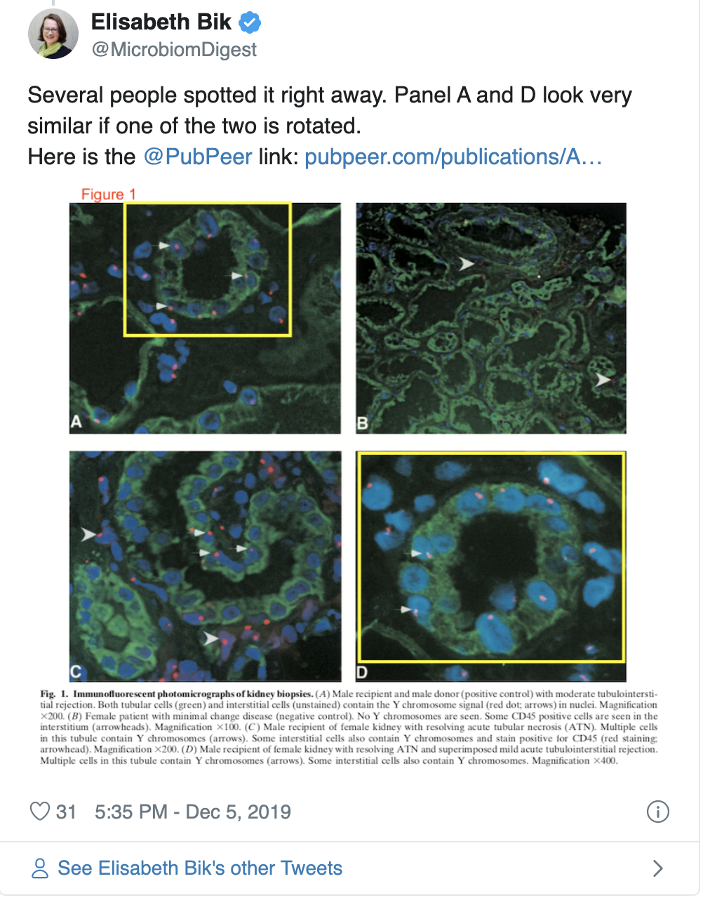
         Elisabeth Bik, a scientist who worked at Stanford University for over a decade, conducts studies on image duplications, a specific type of image manipulation, and brings attention to this issue on her Twitter account. Bik posts images that she suspects have been manipulated on Twitter and often asks others to see if they can spot the duplications that she has found, in a slightly dark game of “I Spy”. Her posts help bring attention to what is becoming an important issue, as both intentional and unintentional image duplications are not uncommon in scientific publishing. A study by Bik, et al. 2016 found approximately 4% of all biomedical publications (sampled across 40 journals) contain some form of image duplication in them, either with unintentional or intentional manipulation, and in a more generalized, self-reported study, roughly 2% of scientists admitted to manipulating data in their work (Fanelli, 2009).

An intensive study published by Fanelli, et al. in 2019 rigorously tested if several parameters contributed to whether or not papers were more likely to contain duplicated images. The study sampled over 8,000 papers published by PLOS One between 2014-2015 and tested if pressure to publish, peer-review, implementing misconduct policies, or gender had any effect on the likelihood of papers to contain manipulated images. The study identified that social control (the level oversight placed on researchers by academic institutions, peers, etc.), cash-based incentives, and whether countries had legal policies in place for image falsifications in academic publishing all contributed to whether image manipulations were likely to occur. In contrast, the author’s gender did not play a role in whether papers were more likely to contain manipulated images. It is important to note that despite image manipulations occurring in noticeable numbers, not all duplications were thought to occur because of an intent to distort data. The types of manipulations identified ranged from unintentional mislabeling to cutting and pasting parts of images, which would be more indicative of an intention to mislead.

The question then becomes, what can be done to identify image manipulations and, ultimately, catch them before they are published? While researchers are actively working to identify duplicated images by eye, others are working to develop software that may be used to catch manipulations before publication. Work by Acuna, et al. 2018 provides a method to detect the duplication of images by comparing images across papers published by the same first or last author. One important limitation of such a method is that it becomes increasingly complex to compare images within a paper to all previously published work; it is why the authors of the method limited the comparisons to works published by the same first or last author. Further, figures can include complex components such as mathematical equations, which may be replicated across publications, or contain objects such as arrows within them that may be duplicated across images. These appropriately duplicated components are mistakenly flagged as image manipulations using the developed software. While these issues present complications when using software that can identify duplication across figures, the authors of this method propose that if journals were to implement the use of such software, that it could deter people from knowingly publishing duplicated images.

In addition to the development of software meant to catch duplications, Fanelli, et al. propose that preventative approaches, such as making and enforcing misconduct policies and promoting research criticism, can help reduce image manipulation in academic research. While this approach rightly aims to change the academic culture that reportedly encourages image manipulation, it will take time for the scientific community to see any effects. In the meantime, what can we be doing to read and think critically of scientific papers, knowing that image manipulation is an existing issue? To keep up to date on the latest in image integrity, and to see what someone with sharp eyes can catch, you can follow Elisabeth Bik on Twitter or look to places such as retraction watch


References: 

  1. Acuna D. E., P. S. Brookes, and K. P. Kording, 2018 Bioscience-scale automated detection of figure element reuse. Biorxiv 269415. https://doi.org/10.1101/269415
  2. Bik E. M., A. Casadevall, and F. C. Fang, 2016 The Prevalence of Inappropriate Image Duplication in Biomedical Research Publications. Mbio 7: e00809-16. https://doi.org/10.1128/mbio.00809-16
  3. Fanelli D., 2009 How Many Scientists Fabricate and Falsify Research? A Systematic Review and Meta-Analysis of Survey Data. Plos One 4: e5738. https://doi.org/10.1371/journal.pone.0005738
  4. Fanelli D., R. Costas, F. C. Fang, A. Casadevall, and E. M. Bik, 2018 Testing Hypotheses on Risk Factors for Scientific Misconduct via Matched-Control Analysis of Papers Containing Problematic Image Duplications. Science and Engineering Ethics 25: 771–789. https://doi.org/10.1007/s11948-018-0023-7 


Additional Articles on Image Duplications:

A recent case of image duplication: https://www.sciencemag.org/news/2019/09/can-you-spot-duplicates-critics-say-these-photos-lionfish-point-fraud

You Eat What You Are

Written by: Sydney Wyatt

Edited by: Hongyan Hao

How nutrigenomics and other genetic information contribute to personalized health in the age of direct-to-consumer genetic testing.

Nutrition has long been touted as a disease-fighting tool. Vitamin C supplements cure scurvy. Diets low in phenylalanine, an amino acid found in protein and some artificial sweeteners, keep phenylketonuria patients’ symptoms at bay. The ketogenic diet was invented to treat epilepsy. However, some of these tools have taken on new, sometimes inaccurate, benefits and companies have exploited these perceived benefits, profiting off of misinformed consumers. A popular example is the use of vitamin C to prevent or cure the common cold. There is no evidence to support this claim, yet the companies behind Airborne and Emergen-C profit off this misguided belief.


That being said, nutrigenetics and nutrigenomics aim to treat and manage genetic diseases like phenylketonuria that rely heavily on dietary adjustments to ease symptoms. However, there is a push to leverage these fields for personal health regardless of disease. With the increase of direct-to-consumer (DTC) genetic testing — 23andMe, AncestryHealth, Orig3n, Helix, etc. — consumers have access to genetic information about their risk of certain diseases and, based on self-reported information on their lifestyle, can get insight as to how these factors play into pre-existing genetic conditions. This information can be powerful, but making changes without a physician’s guidance “may harm the consumer’s health and finances.” Combine this with anecdotal evidence of certain diets managing cancer risk and symptoms, and nutrigenomics can quickly become unreliable at the consumer level. 


Nutrigenomics complements the precision medicine movement by attempting to understand genetic responses to diet and leveraging that information to improve dietary guidelines. Hundreds of studies have attempted to demonstrate gene-lifestyle interactions for obesity and type 2 diabetes, but they had many limitations. For instance, these studies had small sample sizes that were inadequate for statistical analysis and relied on imprecise self-reported dietary and lifestyle data. Recent efforts to replicate the reported findings have failed, therefore the conclusions are unreliable at best. 


A number of factors contribute to health. If these are not properly balanced, there can be negative consequences. This illustrates how important it is to investigate more than just genetics and genomics when creating personalized health plans.


DTC genetic tests using a single locus to evaluate disease risk are not considered medically actionable. As a result, consumers can jeopardize their health by making changes based on this information. Famously, 23andMe received a letter from the US Food and Drug Administration (FDA) in 2013 expressing serious concerns over the results the company provided. The FDA claimed there was too much room for error when testing for diseases like breast cancer using a single locus (BRCA):


“Some of the uses … are particularly concerning, such as assessments for BRCA-related genetic risk and drug responses … because of the potential health consequences that could result from false positive or false negative assessments for high-risk indications such as these. For instance, if the BRCA-related risk assessment for breast or ovarian cancer reports a false positive, it could lead a patient to undergo prophylactic surgery, chemoprevention, intensive screening, or other morbidity-inducing actions, while a false negative could result in a failure to recognize an actual risk that may exist.”


Environmental factors and diet can change the gut microbiome. This can have many effects on distal organs.

The letter continues to explain that consumers may self-manage treatment based on drug response testing with potentially deadly consequences. In the years since, 23andMe has received FDA approval to provide genetic information such as BRCA1/2 breast cancer risk, MUTYH-associated colorectal cancer risk, type 2 diabetes, and Parkinson’s disease. The majority of the test results consist of ancestry and trait reports, which have no medical relevance.


There are too many unknowns for a consumer to DIY a personalized nutrition plan based on their genetic test results alone. However, discussing the results in conjunction with factors such as family medical history and current health status with a physician is a safer way to personalize health. There are other “omics” that can provide more comprehensive information for health management strategies than genomics and nutrigenomics. Metabolomics and gut microbiota analysis offer promising insight into human health. Genetics and the environment can affect metabolism and gut flora composition, so this provides downstream information. Nutrigenomics still has a leg up on these techniques with regards to accessibility and affordability, but there may be a future where these analyses will be part of a routine check-up. Until then, take your test results with a grain of salt and consult your physician before making major changes to your lifestyle.


Bibliography:


  1. Phenylketonuria (PKU). (2018, January 27).
  2. D’Andrea Meira, I., Romão, T. T., Pires do Prado, H. J., Krüger, L. T., Pires, M. E. P., & da Conceição, P. O. (2019, January 29). Ketogenic Diet and Epilepsy: What We Know So Far.
  3. Gunnars, K. (2014, June 17). 20 Mainstream Nutrition Myths (Debunked by Science).
  4. Marshall, M. (2019, October 2). No, Vitamin C won’t cure your cold.
  5. Sommer, C. (2019, June 13). Food as medicine? Scientists are getting closer through nutrigenomics.
  6. Ordovas, J. M., Ferguson, L. R., Tai, E. S., & Mathers, J. C. (2018, June 13). Personalised nutrition and health.
  7. Metcalf, E. (2018, January 5). Photo Gallery: 10 Top Foods to Fight Cancer.
  8. Stern, A. P. (2019, June 17). Feeding the Beast: Could Eating the Right Diet Starve Cancers Like Mine?
  9. Li, S. X., Imamura, F., Ye, Z., Schulze, M. B., Zheng, J., Ardanaz, E., … Wareham, N. J. (2017, July). Interaction between genes and macronutrient intake on the risk of developing type 2 diabetes: systematic review and findings from European Prospective Investigation into Cancer (EPIC)-InterAct.
  10. Guasch-Ferré, M., Dashti, H. S., & Merino, J. (2018, March 1). Nutritional Genomics and Direct-to-Consumer Genetic Testing: An Overview.
  11. Dickey, M. R. (2013, November 25). The FDA Wants 23andMe To Stop Marketing Its Genetic Testing Kits.
  12. McNiven, E. M. S., German, J. B., & Slupsky, C. M. (2011, October 12). Analytical metabolomics: nutritional opportunities for personalized health.
  13. Cheng, M., Cao, L., & Ning, K. (2019, October 9). Microbiome Big-Data Mining and Applications Using Single-Cell Technologies and Metagenomics Approaches Toward Precision Medicine.

It’s not just a phase (separation)

Written by: Jennifer Baily

Edited by: Sharon Lee


Phase separation is the process by which one homogenous mixture separates into two distinct phases. Imagine trying to shake a bottle of oil and water. The two solutions mix initially, but then separate into little spheres, eventually coalescing into two separate phases: one made up of oil and the other of water.

Graphic Source

This sounds obvious, but how it occurs in cells, its regulation, and methods used to investigate this phenomenon are infinitely more complex. Not surprisingly, phase separation is a key regulatory mechanism in biology. Membraneless organelles such as P bodies, nucleolus, and germ granules are commonly formed through liquid-liquid phase separation, a type of protein phase separation. This further compartmentalizes the cytoplasm alongside lipid-bilayer organelles like the mitochondria and nucleus. Typically, macromolecules must exceed their solubility limit to permit liquid-liquid phase separation (PS). Macromolecules then condense into droplets and distinctly separate from the dilute surrounding phase. 

Since the discovery of the nucleolus in the 1830s, we have learned that liquid-liquid PS contributes to the formation of many intracellular compartments. Phase separation may also contribute to the formation of subcellular structures, such as heterochromatin, as well as membrane receptor clustering. The key functional aspect of these phase separations are the compartmentalization of cellular structures without the need for lipid bilayers.

Through proteomic and genetic screens, several crucial properties have been identified in the formation of these phase-separated particles. Multivalency and linear motifs are often found in proteins undergoing phase separation. This multivalency allows for “well defined interaction surfaces… which can participate in stereospecific interactions”. Intrinsically disordered regions of proteins may permit association by interacting in a variety of ways to drive phase transitions. Research suggests they work primarily through “sticky” interactions facilitating assembly due to “the exchange of macromolecule-water interactions for macromolecule/macromolecule and water/water interactions under conditions for which this process is energetically favorable”.

Graphic Source

Interestingly, membraneless organelles can be liquids, solids, or gels. These properties are dictated by spatial ordering, which is determined by the organelle length and macromolecule organization. Liquid phase separated droplets display typical behaviors of liquids – fusion, dripping, etc. – and all properties are determined by the separated material’s surface tension. Unlike their more dense hydrogel counterparts,  liquid phase separation is often reversible. The formation of solid phase separated states is thought to be due to protein sequences; however, even gel and solid phase separated biomolecular condensates have displayed dynamic behavior. The dynamics of liquid-liquid phase separation are thought to play a key role in the condensates’ function, whether it be segregation, assembly, activation, inactivation, or localization. One Biochemistry, Molecular, Cellular, and Developmental Biology graduate student at UC Davis studying phase separation described the moment when they “unexpectedly observed the dynamic liquid droplets forming from the cells”, and it completely blew their mind. After studying cells using static methods such as fixation, staining, and immunofluorescence, they were surprised to find these dynamically forming lipid droplets while live imaging.

While phase separation has been shown to be an integral part of cellular processes and illustrates the complexity and beauty of biology, some scientists still question its legitimacy. Whenever a new process is shown to have phase separation properties, it is important to consider the possibility that protein overexpression can induce aggregation, imaging confounds, and other artifacts that resemble phase separation characteristics but do not represent endogenous properties. I think the BMCDB student sums it up best: 

“The notion that liquid-liquid phase separation may be a general mechanism by which cells use to concentrate specific molecules and to facilitate the formation of membraneless organelles is compelling. However, the evidence for phase separation in vivo, especially in its endogenous abundance and physiological condition, is scarce. In addition, instead of comprehensive quantitative measurements, most studies to date have used qualitative evidence (i.e. roundness, fusion and splitting events of droplets) to demonstrate phase separation. Furthermore, the functional consequences of phase separation in vivo remain to be determined, given that perturbing phase separation property specifically without affecting other functions of the protein is still challenging to do. Therefore, better assays and more advanced techniques are needed to advance the field and to reveal true biological insights of phase separation in vivo.”

Plastic use in basic science

Written By: Mikaela Louie

Edited By: Keith Fraga


Despite growing public awareness of its impacts, plastic use continues to have detrimental effects on the environment. Public opinion turning has pushed companies to use biodegradable/compostable packing (i.e., eco-friendly materials) and to invest in new sustainable practices. One recent example is Starbucks’s plan to stop using plastic straws by 2020. 

Industry development of biodegradable plastics is also growing, but the types of products available are limited. Additionally, the vague definitions of biodegradable, compostable, and recyclable plastics are not transparent to the public and are often used to “green wash” products that might otherwise continue to have a negative impact. For example, Peak Products has developed biodegradable nitrile gloves. While this is a promising step towards sustainability, it is unclear if these gloves actually degrade faster in landfills than regular nitrile gloves and that the products resulting from their breakdown are indeed better for the environment. As a biology graduate student researcher, I often find my research practices in conflict with my concern for the environment. The need for sterile materials at the lab bench results in prolific use of single-use plastics. Therefore, while many industries work to ban plastic use, most research facilities cannot comply with these changes and are left with few options for alternative materials.


Environmental Impact of Plastic

Plastic waste is mounting in the environment. An estimated 8300 million metric tons (Mt) of plastics have been manufactured to date, resulting in 6300 Mt of plastic waste, the most common being plastic water bottles. Only 9% of this waste is recycled, with 79% of it heading to landfills and the remaining 12% being incinerated. Macroplastics, such as plastic water bottles and disposable utensils, disposed in landfills end up in agricultural land and the ocean as microplastics. Consumption of these microplastics by marine organisms has a severe impact on the marine food chain due to the physiological damage caused by ingestion. Not only are we destroying the surrounding environment, but we are also disrupting the health of our food sources.

The most commonly used plastic for single-use items is polyethylene, and thankfully there is evidence of bacteria and fungi that degrade polyethylene. However, the landfill environment is most likely not conducive for the degradation of such large amounts of waste by a small population of microorganisms. 

In an average week (7 days because the cells won’t let me have weekends), I use more single-use plastic than I care to admit. I broke down the amount I use by experiment and type of item: 


Cell culture maintenance (one plate)

Volumetric pipette: 14

Glass pipette (not plastic but still trash): 14


Passaging cells (usually 2x a week)

Pipette tip: 16

Volumetric pipette: 12

15mL tube: 4


qPCR (4 different samples, 5 targets) 

Pipette tip: 82

1.5mL tube: 10

qPCR plate: 1


Immunohistochemistry on tissue sections

1.5mL tube: 5

Pipette tip: 16

Figure 1: Visual representation of average plastic use in one week.

These averages are also assuming that I don’t make any mistakes, forcing me to throw the item away without actually using it. Even if I made zero mistakes, this amount of plastic from a single week would take thousands of years to degrade. Reflecting on my plastic use and its environmental impact has motivated me to learn what changes are being made and to help align many researchers’ environmental values with their research practices, including my own.


Green Lab Programs

Many universities have begun their own green lab programs, such as MIT and here at UC Davis. Most of these programs evaluate labs on several categories, including energy use, waste production/management and transportation. Labs can participate in these programs to gain certification and to contribute to university wide sustainability practices. 

Outside of university-run programs are non-profit groups like My Green Lab. They have created several programs, including a Green Lab certification and the International Laboratory Freezer Challenge. The Freezer Challenge is a competition to see how efficiently labs can store samples and maintain freezers to reduce energy output. The Green Lab website also includes several links to resources for ways labs can implement sustainable practices, such as glove recycling and waste reduction tips. Vendors also have recycling programs for items such as gloves, empty plastic packaging, and pipette boxes. Origin Materials, a UC Davis alumni founded start-up, is also developing biomass polymer products that can supposedly degrade faster than petroleum based plastics. Most of these programs require effort from labs to sort recyclables, and usually only accept items that have no hazardous contaminants. These programs can be established for labs by contacting the local vendor representative. 


A Few Takeaways 

Single-use plastics are extremely useful. They have allowed science to design well-controlled experiments in an economical way. However, we cannot ignore the consequences that we now face due to our enthusiastic plastic use. It is undeniable that we cannot return to a culture without plastic, but it is becoming more and more necessary to develop plastics that more readily degrade and/or better recycling methods than the ones currently in place. We are also entering a new era of recycling due to China’s refusal of our recyclable waste. This poses an urgent need for better recycling infrastructure within the US. How will this change the ecosystem of materials, packaging, and consumer marketing? Will there be a cultural shift to better recycling practices, such as infrastructure to process difficult-to-recycle materials, and commitment to necessary education for proper recycling practices? 

Lessening the amount of plastic that we use is probably the best way that we can reduce our carbon footprint in labs without establishing new programs. This is pretty hard to do with the need for sterility, yet can be accomplished by planning experiments ahead of time and minimizing mistakes. In 2016, UC Davis was named the most sustainable university in the world. Therefore, it seems only fitting that we apply sustainable practices in our research labs as students of UC Davis. 

My experiences navigating mental illness as a graduate student  

Written By: Jennifer Baily

Edited By: Emily Davis

Trigger warning: references to suicide


I was diagnosed with depression during my sophomore year of college, but I also remember an instance where at 12 years old, I prayed for the strength to end my own life; this was not an unfamiliar feeling. I was lucky enough to be well-supported during several life-changing experiences throughout adolescence and college, thanks to friends, family, and undergraduate student resources. What I didn’t know was the extent to which my mood disorder would affect my graduate experience, whose signs and symptoms were never communicated to me by mental health professionals.

For 3 months during my second year of graduate school, I was convinced that I had mononucleosis. I was exhausted and drinking two energy drinks a day, to the point where it became a joke amongst my lab mates. I was tested for everything you can think of, including the suspected mono, B12 levels, and even thyroid disease. My doctor recognized the exhaustion, lack of appetite, and social isolation for what it was – the beginning of a major depressive episode.

Instead of agreeing to increasing my dose of antidepressants, I was convinced that it was stress related to an upcoming qualifying exam, 3 months in the future. Despite sincerely believing that I was fine, my doctor heavily encouraged me to meet with her every two weeks to monitor my health. A fellow graduate student suggested that I start counseling again, which I did at the Student Health Center on campus. Counseling helped to a certain extent. We talked about how to strategize getting through difficult situations and conversations, and how to practice self-care. 

Unfortunately, the on campus clinic was unable to provide adequate care because I didn’t know how necessary it was to communicate what was going on internally; until recently, I was unaware that my near-constant suicidal thoughts were not a part of the day to day human experience. My mental illness manifests itself through using work, reading, and planning as coping mechanisms to avoid sitting with these thoughts. In my experience, high-functioning depression flies under the radar in an environment where many are working long hours with few results.

I lost ten pounds in a month. With concerted effort, I gained most of it back, but my bones always seemed to hurt with a dull ache. During a major depressive episode, the brain’s pain threshold is lowered – normal aches and pains become distracting to the point of affecting your ability to function. I didn’t know that one side effect of extreme depression was a state called “pseudo-dementia”, where the patient cannot form long term memories; I certainly struggled recalling basic facts about myself, even more so those which I had studied in preparation for my qualifying exam.

The exam itself, and 8 months preceding, are a blur of exhaustion that I can barely recall. I knew that I had not presented my best work, and was disappointed that the months of rigorous preparation did not support me during current mental state. I spiraled. A month later, a good friend came by to see if I wanted to grab coffee. I had been staring at my computer, desperately trying to make sense of the jumble of words before me, immediately forgetting sentences after discovering their meaning. 

In chatting with her, I opened up about how living through the week, even more the rest of my life, was exhausting to even think about. I seemingly always had thoughts, but now I had a plan. That day, when I was already at my breaking point, several minor instances brought me to my knees. My friends tried to explain the facts of the situation for what it was, something that can easily be overcome. I was beyond thoughts and plans. I told my friends over text that I should just kill myself, and I believed it. 

In that moment, I didn’t recognize myself, my life, or anything valuable. When you struggle, refusing assistance for nearly 6 months, you reach a level of pain and suffering that is absolutely crushing. I will not go into detail further because that is not the point of this article. What I will say is that without incredible friends supporting me in a time of great need, I would not be here. I don’t know if I can adequately thank them.

The next morning, I sent a message to my doctor explaining the situation and finally agreeing that there was a problem and to up the dose of my antidepressants. I was not in my right mind, thinking that this was an adequate solution. If you or anyone you know feels this way, immediate professional help is absolutely required. The doctor called me repeatedly, and made me an appointment with the on-call psychiatrist, despite me arguing that I had a really important time course experiment, which I – in that moment – valued more than my own life.

Needless to say, I got the help that I needed. My PI was incredibly supportive about my hospitalization, giving me the time that I needed to get better. Normally, involuntary hospitalization would be the route, but because I didn’t cause any actual harm (thank you friends who came over), the on-call psychiatrist placed me in a partial hospitalization program (PHP). Consenting to voluntary hospitalization was the most difficult and humbling experience of my life so far. In a PHP, as opposed to involuntary, you get to go home at night and on the weekends, after going over a safety crisis plan with your provider. I am so grateful that my student health insurance plan covered the ~$24,000 of care.

I was picked up by an unmarked, white van the following morning, and was taken to a nearby hospital. There I met amazing and diverse people struggling with the same illness. For the first time, I felt understood, albeit a little crazy. I was overjoyed to eat slimy hospital-cooked fish because I hadn’t been able to feed myself in a few days. I was given medication and finally slept after getting a cumulative three hours of sleep over the preceding three days. During my time in a partial hospitalization program, we learned more about the biology of our illnesses, and which coping skills could help us in the situations that brought us there in the first place. I returned to work, with some hitches, after being transferred to an intensive outpatient program where I attended a shorter series of therapy and workshops.

Some people thought I had taken a vacation, while others had heard rumors that I had begun a planned leave program. Coming back, I was wracked with shame and guilt. I thought that once I fixed myself, all my other problems with school, lab work, and personal relationships would be fixed as well. My release from the hospital did not mark an end to the fragility of my situation. I struggled with the idea that I was more inferior than I already felt because of my mood disorder (imposter syndrome anyone?). I watched a horror movie with a suicide scene, was unable to control my tears in the theater, and didn’t sleep for a few days. I still had trouble focusing, making reading even brief reviews incredibly difficult. Sometimes I still think that it would be easier to simply disappear. But I know that these thoughts will pass and do not influence how I choose to live my life. 

All this to say, if you are or if you have a graduate student struggling with mental illness, you are not alone. Based on statistics provided by the student health center at UC Davis, “15.5% of undergraduates seriously considered suicide (3.6% increase since 2015), 2.6% had attempted, nearly doubling  since 2015’s 1.5%, and 8% of graduate students reported seriously considering suicide”. 

I want this article to encourage those who are in a similar situation that, with help, things will get better, to help mentors who may not understand the level to which their mentees may be affected, and to validate the experience of other graduate students struggling with mental illness. Not only are you not alone, but there are many people and resources ready to help, so long as you take advantage of them. Some of these resources I didn’t know about until after being hospitalized, and I continue to learn about more as I open up about my experience.


On campus services – UCD specific

https://leadership.ucdavis.edu/strategic-plan/task-forces/mental-health

  • Individual counseling
  • Couples counseling
  • Group therapy
  • Skills groups
  • Case management
  • Career counseling
  • Outreach to the campus community

They only work if you use them.


Partial Hospitalization and Intensive Outpatient Programs

Group therapy at a hospital location, often using cognitive behavioral therapy, with access to on-call psychiatrist and nurse.


National Suicide Prevention Lifeline

We can all help prevent suicide. The Lifeline provides 24/7, free and confidential support for people in distress, prevention and crisis resources for you or your loved ones, and best practices for professionals.

Online Chat – Lifeline

1-800-273-8255


“Warm Line” – Talk to trained listeners before a crisis

Visit http://www.warmline.org/ to find the number for your area.

Psychedelics in Biology and Mental Health 

Written by Keith Fraga

Edited by Mikaela Louie


Psychedelic drugs are often taboo in US culture particularly since their extensive criminalization in 1970. Substances like LSD, MDMA, DMT, and those derived from fungi like psilocybin, are drugs known to have hallucinogenic effects on humans. The ability of these substances to produce mind-altering experiences lead to massive efforts to control their study and use.

However, restrictive policies surrounding psychoactive and other mind-altering substances may be changing. For example, cannabis is undergoing rapid social and legal acceptance in the US. The City of Oakland legalized the recreational use of magic mushrooms in June, another potent hallucinogenic. Substances such as ayahuasca have a long history as a traditional medicine. Whatever impact psychedelics have on treating debilitating mental illnesses deserves experimental study and mechanistic understanding. Indeed, psychedelics are in several clinical trials to treat various mental illnesses, like depression, anxiety, PTSD, with the potential to be applied to many more mental health conditions. UC Davis researchers recently published an article in Cell on a study of psychedelics’ effects on neural plasticity.  

Diving into the literature, I wanted to answer a few specific questions. What is the history of psychedelic research? Where did restrictive policies surrounding psychedelics come from in the first place? What has contributed to the thawing of restrictions on clinical psychedelic use ? How has biology and neuroscience changed our understanding of these substances? And lastly, what do we not know and where should we be cautious?


HISTORY

The history of psychedelic drugs help us understand their possible future. Psychedelics refer to many substances, and naturally occurring psychedelics such as psilocybin or ayahuasca have been consumed by humans for centuries. Others are more recent in human history, being chemically synthesized in the late/early 19th-20th centuries. When consumed, psychedelics generally cause an altered state of perception. Often these altered states affect tethers to reality, mirroring what happens in psychosis. The history and story of lysergic acid diethylamide (LSD) is an important example of the use and study of psychedelic drugs.

LSD was first synthesized by Swiss scientist Albert Hofmann in 1938. Hofmann set out to synthesize compounds that could be used for blood vessel constriction, which could help in medical applications to reduce blood loss. After accidental contact with LSD, Hofmann found that this compound had hallucinogenic properties. 

Hofmann self-administered doses of LSD to test and observe its effects, becoming convinced of LSD’s potency to address mental illness. Hofmann contributed to the spread of LSD from doctor to doctor, making its way to the US. It was quickly found that LSD had potent therapeutic effects for treating various psychological conditions and became wildly presubscribed by therapists across the US and Europe.


USE AND REGULATION IN THE UNITED STATES

An important aspect to the rise in LSD usage was the prescription and research by independent, unregulated clinics and therapists. In this unregulated climate, LSD found its way out of the laboratory and into the 1960s US counter-culture. The culture around psychedelic research and usage at this time was very exploratory. As with the initial discovery and subsequent spread of LSD, therapists and researchers self-experimented and shared these compounds without discretion.

The obvious potent effects of LSD spurred a generation’s worth of compelling, rigorous research. Figure 1, shown below, depicts the relative citation rate for the terms “psychedelic” or “lysergic acid diethylamide” in any journal article title/abstract. The years between 1950-1970 can be called the “golden-age” of clinical and basic research on uses for LSD. During this period, much was learned about LSD’s effects on human psychology, crucial observations on the aspects of psychedelic “trips”, effective uses of LSD in psychedelic assisted therapy, all of which are now being re-examined with modern medical and biological methods.


Figure 1: Citation rate for words “psychedelic” or “Lysergeic acid diethylamide” in journal article title/abstracts. The data series in blue is just for “psychedelic” or “Lysergeic acid diethylamide”, while the red series is a separate search with additional search terms for other psychedelic substances. Find the interactive version here.


It is also clear from Figure 1 is the “golden-age” of LSD research rapidly came to an end. In response to growing concerns for unregulated, unethical use of LSD, the Food and Drug Administration adopted new policies restricting the use, possession, and manufacturing of these drugs. A series of legal policies were installed in the ‘60s to curtail the use, possession, manufacturing, and research into these compounds, culminating in the Controlled Substances Act (CSA) in 1970. The CSA is the legal authority that established the Schedule system for controlled substances. Schedule I is the most restrictive category of compounds. With strict policies and massive legal consequences for inappropriate use of these substances, many researchers and therapists in the field ceased their work.


THAWING OF RESTRICTIONS ON PSYCHEDELIC RESEARCH 

It is difficult to identify a singular cause for the increase in interest and research on psychedelics. That being said, I find two specific developments particularly informative to the growing acceptance of psychedelic research. First is the research on how psychedelics can improve the quality of life for patients with life-threatening disease or terminal illness. Patients with life-threatening diseases, terminal illness, and associated chronic pain experience lower quality of life, higher rates of depression, and lower life-expectancy. While researching for this article, I found the following interview with Michael Pollan, a journalist who studies the impacts of psychedelics and their potential growing role in society. In this interview, he relates what sparked his interest in therapeutic use of psychedelics was their ability to improve the mental health of terminally ill patients. Numerous recent studies and reviews have recently contributed to this area of research. Psychedelics in end-of-life care was actually studied in the 60’s, and it is now being revived. Current research on therapeutic effects of psychedelics has demonstrated they can dispel anxiety, fear, and despair in these patients safely, and social taboos and legal policies are slowly changing in order to increase access and study of these benefits.

Psychedelic assisted therapy is also being explored for an array of mental health conditions. Numerous psychotherapy drug trials currently ongoing. The MultiDisciplinary Association of Psychedelic Studies (MAPS) is a central scientific research coalition propeling psychotherapy trials forward. MAPS independently supports trials for psychotherapy with a wide array of substances to tackle a diverse set of mental health conditions. Specifically, MAPS is currently working with the FDA for MDMA assisted psycho-therapy.   

Psychedelic assisted psychotherapy presents an alternative therapy regimen for mental illness in part because of the global effects psychedelics have in the brain. A significant hypothesis in this field is psychedelics and psychiatric medications address mental illness at different scales. Psychiatric medications, like antidepressants, offer to rectify chemical and functional imbalances in the brain. These often target specific neuro-transmitter/receptor imbalances. However, psychedelics impact brain activity at a systems level, disrupting default neural networks and activating connections between disparate regions of the brain. The golden-age of psychedelic research in ‘60s intuitively reached many of these ideas concerning the global effects in the brain psychedelics have. With modern developments in science, medicine, and biology, researchers can interrogate psychedelic compounds’ effects on brain function at greater resolution. 


A BIOLOGICAL MAGNIFYING GLASS TO PSYCHEDELICS

A significant motivation of this article is a study by a team of UC Davis scientists studying the effects that a battery of psychedelic drugs have on structure and dynamics of neurons. This study experimentally demonstrated that many psychedelic compounds promote neurons to make new synapses, growth of new neuron axons, and altered neurophysiological functionality. In part, this is why psychedelic compounds are termed by the authors of this study “psychoplastogens” for their ability to promote plasticity at the level of the brain. These investigators also experimentally tested mechanistic models for how these large-scale structural and functional changes in response to psychedelics. Investigations similar to the UC Davis study represent how advanced techniques in biology can garner quantitative insights into the mode of action for these potent compounds. 

A greater understanding of how psychedelics functionally work in the brain can lead to the generation of new compounds that have similar functional effects demonstrated in the above study, with the idea being these newly designed compounds may induce similar subjective experiences, or “trips”. In other words, the generation of novel “psychoplastogens” is possible when we learn more about what determines their function in the brain. Alternatively, psychoplastogens can be used to perturb aspects of the brain to piece together the mechanisms for subjective experience. Biology has a rich tradition of using tools to perturb a biological system and measuring its output to uncover the mechanisms that make it work in the first place. Psychedelics can serve a similar role in potentially understanding perception, cognition, and consciousness.    


CAUTIONARY TALES

Nearly every editorial, podcast, or interview I have heard on this subject includes strong cautionary tales. Psychoactive drugs can have unintended consequences.The mind-altering capabilities for these drugs are so potent that they may do more harm than good for some people, which is one reason why I am particularly excited about the prospect of trained therapists guiding a patient’s trip. How the chemistry of these molecules and their therapeutic use come together to help people is an exciting new venture in medicine.


Page 3 of 3

Powered by WordPress & Theme by Anders Norén