A UC Davis Graduate Student Blog

Author: Staff Page 2 of 3

Mastering the art and science of good presentation design

Powerpoint presentation is one of the most effective ways to communicate our science. However, mastering the art of presentation can be a long and enduring journey. There are two parts of a good presentation. One is about speaking, and the other is the actual presentation itself. If you have a fear of public speaking, please check out our previous post on “combat the public speaking fear”.

This post focuses on the design components of a good presentation. Luckily for us, there are many fundamental principles that you can learn from this short-ish video to help you achieve that mastery. This video, suggested by Hongyang Hao from Dan Starr’s lab, features Stanford neurologist Susan McConnell. Susan McConnell is a world-respected scholar and science communicator with iBiology. She is a member of both National Academy of Sciences and the American Academy of Arts and Sciences, and she is also both an HHMI scholar and a Pew Scholar. The list goes on and on.

You may know some of the recommendations already from the video, especially, if you are a UC Davis BMCDB graduate student who went through our awesome rotation class. However, I’m sure there is much more new information that you can learn from this video. Also, it is just a good practice to systematically evaluate your presentation skills once a while, and make sure your presentations don’t have the pitfalls mentioned in the video.

Mentioning the rotation class made me remember my first presentation in that class. All I can say is “ughhhhh”. I’m so glad it is better now.  

Yulong

Want to see more awesome videos like this one? Make sure to check out iBiology and their videos on the importance of giving a good presentation and more tips on creating effective slides.


Suggested by Hongyan Hao

Edited by Anna Feitzinger, Keith Fraga

For any content suggestions or general recommendations, please email to UCDBioScope@gmail.com and put science 2.0 in the title.

Do Regulations on Genomic Data Inform or Mislead the Public?

Author: Emily Cartwright

Editor: Hongyan Hao


As sequencing costs drop and more companies develop genomic testing for everything from predicting hair color to the risk of disease susceptibility, the public has access to a wealth of personalized health information. At the same time, companies are faced with the decision of how much information to release to the public, only some of which is regulated. The Food and Drug Administration (FDA) has policies on what kinds of information that companies such as 23andMe and Ancestry.com can release to the public, but there are many enterprises that are not clinically certified and utilize genomic data to conduct health studies (1). How to regulate and disseminate findings from these companies, especially smaller scale studies that may not be clinically certified, is an unresolved issue.

Many of the current FDA regulations for companies like 23andMe are disease specific. Regulations released in 2017 allowed 23andMe to report on variants associated with 10 diseases or conditions (2) and in 2018, allowed the release of information on three BRCA1/BRCA2 variants to consumers who submitted for genetic testing (3). These specific BRCA1/2 variants are known to be associated with an increased risk of developing cancer but there are over 1,000 identified BRCA mutations (3). While these regulations limit the amount of information that consumers are provided, they also circumvent the current issue of how to relay health information to patients in a truthful and informative way; where it is made clear that the results of genetic testing indicate the known risk of developing a certain condition.

It is crucial that companies emphasize that not having a genetic marker associated with a disease or condition does not mean that the individual will not develop the condition. Current regulations from the FDA seek to limit what information can be released to the public but do not address how large companies can best convey health information to consumers. There are also gaps in the ability of agencies like the FDA to regulate all health information gleaned from genomic data, as many small companies do not fall under their regulatory jurisdiction (1).

Recently, the National Academies of Sciences, Engineering, and Medicine (NASEM) released a report (4) encouraging researchers to relate findings from biological data analysis back to people whose samples were used in the studies. The report is intended to increase the flow of information from scientists to the public, but a central issue still involves the question of how to convey information in a way that is both meaningful and accurate, in terms of whether or not a finding has medical significance

Genetic variants may indicate risk of disease susceptibility, but this is not necessarily causal and relaying such information to the public can be tricky (1). Researchers are also faced with an increased cost of funding for these studies because determining how to relay information to the public and executing this can result in additional expense (1). The NASEM recommended that researchers should plan out what information they will release from their studies prior to starting them, which may also help the researchers to plan out the added cost of relaying this information (1,4).

The flow of information from researchers to consumers is important but the fact that many studies are carried out by third parties also lies at the forefront of this issue. Companies that do not do the initial genomic sequencing and that may not have the clinical or scientific background to make the diagnoses they relay to consumers may give misinformation (1). The New York Times reported on a case where a doctor had sent his genomic data to a third party, Promethease, and was told that he had a variant known to be associated with Lynch syndrome, a disease that predisposes individuals to cancer early in life (5). The variant was associated with the disease but was not known to cause it, and more importantly, after sending out for genetic testing at a medical diagnostic firm, the doctor received results confirming that he did not actually have the mutation (5). The discrepancy in results brings up the issue of who should be allowed to handle genomic data and how should companies that do not fall under the umbrella of FDA regulations be treated (1). Right now, there may not be a good way to handle third party testing centers that have not been clinically certified, but consumers should be aware of the potential misinformation in results from these companies.

While there are still grey areas concerning the regulation of findings from studies that utilize genomic information, there is an effort to move towards defining what should be released and how. As researchers move towards utilizing this type of data for health and genetic studies, organizations such as NASEM are becoming more critical to help define what and how this information is treated and disseminated. There are many third-party companies that will not fall under guidelines set out by the NASEM but there is still a need to regulate how and what these companies can do with genomic data as well as how their information is released to the public (1). This can come from both sides, in conveying more accurate and meaningful data to consumers and in being more direct about the difference between association and causality in the relationship of variants to disease or condition.


Works Cited:

  1. Couzin-Frankel, J. (2018). If you give your DNA and tissues to science, should you get a peek at what they might contain? Science (New York, N.Y.). http://doi.org/10.1126/science.aau7323
  2. Administration, U. F. A. D. (2017). FDA allows marketing of first direct-to-consumer tests that provide genetic risk information for certain conditions.
  3. Administration, U. F. A. D. (2017). FDA authorizes, with special controls, direct-to-consumer test that reports three mutations in the BRCA breast cancer genes.
  4. National Academies of Sciences, Engineering, and Medicine, Health and Medicine Division, Board on Health Sciences Policy, Committee on the Return of Individual-Specific Research Results Generated in Research Laboratories, Downey, A. S., Busta, E. R., et al. (2018). Returning Individual Research Results to Participants: Guidance for a New Research Paradigm. http://doi.org/10.17226/25094
  5. Kolata, G. (2018, July 2). The Online Gene Test Finds a Dangerous Mutation. It May Well Be Wrong. The New York Times. Retreived from https://www.nytimes.com/2018/07/02/health/gene-testing-disease-nyt.html


Additional Links:

Spit is a podcast put out by 23andMe and iHeartRadio that features celebrity dialogue, highlighting some of the social issues surrounding genomic data.

On privacy issues surrounding genomic data: https://www.theverge.com/2018/8/1/17638680/genetic-data-privacy-consumer-rights-guidelines-23andme-ancestry

And in the news: https://www.nytimes.com/2018/10/18/opinion/sunday/dna-elizabeth-warren.html

More on the role of NASEM in regulating the release of information from genomic studies: https://www.biospace.com/article/national-academies-report-provide-patients-with-clinical-study-results/

Ain’t nobody got time for bad graphs

The above graphs were made from data of an anonymous 4th-year graduate student’s drinking habit, as he became an alcoholic in denial. Although the graphs were made from the exact same data, it’s obvious they give different perceptions of what the data is representing. One of the graphs indicates an alcoholic graduate student slowly increasing its alcohol consumption. The other graph suggests a more destructive trend: sobriety to a full-blown alcoholic. Bad data representation can be just as detrimental as bad data. Anna Feitzinger from the Lott Lab sent me this very useful website discussing common problems faced when graphing data. It has example problems, clear solutions, and some even have convenient practice R code.

https://www.data-to-viz.com/caveats.html


Yulong  


Suggested by Anna Feitzinger

Edited by Sydney Wyatt

For any content suggestions or general recommendations, please email to UCDBioScope@gmail.com and put science 2.0 in the title.

The Art in Science and Science in Art

Author: Anna Feitzinger

Editors: Hongyan Hao, Sharon Lee, Keith Fraga


At first glance, science and art may appear to be two areas of human activity that could not be more distantly related. I’d like to dismantle this idea of scientists and artists and explore the qualities shared by both. Neon color tube racks and primary-colored pipette tip boxes are some of the basics that are scattered about the biologist’s benchtop. Biologists often visualize the microscopic world using dyes or fluorescent molecules and lasers of various wavelengths of light. The result is an image (or video) of colorful molecules whose arrangement depends on the particular cellular landscape and whose interpretation is up to the scientist. In fact, the first image that surfaces when searching “immunostaining” resembles that of a Paul Jackson Pollock painting (Figure 1).

Figure 1.  Left: Immunostained section of brain tumor in Wikipedia, The Free Encyclopedia. Right: Number 1, 1949 by Jackson Pollock. 

The history of science is full of art. Before cameras, scientists had to draw out their discoveries as lens’ became better at high levels of magnification. Inspired by the drawings in Robert Hooke’s Micrographia, the tradesman Antony van Leeuwenhoek learned how to grind lenses so to take up the hobby of microscopy. He made the most powerful lenses of the time and consequently discovered bacteria, protists, sperm cells, blood cells, rotifers and more before his death in 1723. [1] Although he had no formal higher education, he is now considered the Father of Microbiology.

Another unlikely scientist, Santiago Ramón y Cajal, was a rebellious and anti-authoritarian youngster who was said to have been imprisoned at the age of eleven in 1863 for destruction using a homemade cannon [2]. An avid painter, Santiago’s illustrations of brain and neural anatomy have become iconic. Considered the Father of Modern Neuroscience he postulated the neuron theory; the law of the dynamic polarization of the neuron and discovered the axonal growth cone.

Figure 2: A depiction of neurons in the cerebellum by Santiago Ramón y Cajal

Visual artists confined to physical media must discover the nature of their medium. Different techniques require different media and skills. For instance, one must decide the dilution factor of oil paint to linseed oil or acrylic paint to water in order to create a preferred texture and consistency. Screen printers and photographers must learn the properties of the light sensitive emulsions and films with which they work. Musicians become familiar with the sound waves they produce and modulate. Much like scientists mixing reagents in the lab, artists have an array of media used for experimentation.

Scientists and artists both have a need to share their perceptions of the universe which drives dedication, patience and persistence. Both have visions which they spend years perfecting and require creativity for success. Both get feedback from their peers and share their work with the community. Scientists share their work at conferences and artists do so in galleries or on stage. Occasionally, these two worlds collide. Last year marks the twenty year anniversary of the “Worm Art Show” which was started in 1997 by Ahna Skop at the International C. elegans conference. Works presented at the very first show included the C. elegans genome sandblasted onto a piece of driftwood and a wooden construction of the C.elegans vulva [3]. The American Society of Microbiology now hosts an annual Agar Art competition. This years first place piece, “The battle of winter and spring” features a stunning portrait of two contrasting figures made using colored microbes resistant to different antibiotics on a plate of agar.

Artists and scientists share a common thread historically, and each rely on one another in the modern world. Some recent collaborations include the turning of data into sound, called sonification. Mark Ballorad of Pennsylvania State University in State College has received two $50,000 grants for a project aiding marine biologists in translating data from the deep ocean into sound. Being able to listen to large data sets allows for an entirely different perspective that may not be captured by more conventional visualization methods. A recent publication by a music professor and chemical biologist explore how turning protein sequence and structural information into melodies can facilitate analysis.

Here at UC Davis the Department of Design hosts the LASER Series: conversations in art and science. Each LASER event has four presentations from four different disciplines as unrelated as possible. These talks are meant to facilitate interdisciplinary conversations between artists, scientists and generalists. The seminar is part of an international speaker series, all which are documented and accessible online. Another Art/Science initiative on campus is the Art/Science Fusion program which aims to bring creative energies from the arts and sciences to foster innovation.


[1] Kruif PD (1926) Microbe Hunters. Transactions of the American Microscopical Society 45(3):259.

[2] McMenemey, W. H., M.D. (1952). Section of Pathology (Vol. 46). Proceedings of the Royal Society of Medicine.

For updated information on the LASER series at UCD: https://www.facebook.com/laser.ucd

Additional articles on this topic:

https://www.forbes.com/sites/quora/2016/03/16/why-art-and-science-are-more-closely-related-than-you-think/

https://www.ebsco.com/blog/article/the-steamy-relationship-between-art-and-science

https://cosmosmagazine.com/society/when-arts-and-science-collide


Combat the fear of public speaking

What’s your top 5 biggest fears? Is public speaking one of them? A study in 2014 from Chapman University concluded public speaking is one of the top 5 fears faced by Americans. So, if that’s one of your top fears, you are not alone. To help you combat that fear, here is an excellent video from Stanford Graduate School of Business. There are many useful tips and tricks in this very engaging video that can help you to diminish the fear for public speaking.  For example, by changing the style of the talk to more conversational, it could really lower that stress level.


Yulong


Suggested by Hongyang Hao

Edited by Anna Feitzinger

For any content suggestions or general recommendations, please email to UCDBioScope@gmail.com and put science 2.0 in the title.


Microtubule motors read MAPs

Author: Hongyan Hao

Editors: Anna Feitzinger | Emily Cartwright, Keith Fraga, Jessica Huang, Sharon Lee, Yulong Liu, Linda Ma


Long distance cargo transport in neurons is facilitated by microtubule motors kinesin and dynein. In vivo, microtubules are crowded environments covered by different kinds of microtubule associated proteins (MAPs). How do motors react when they encounter MAPs? A recent work entitled ‘competition between microtubule-associated proteins directs motor transport’ published in Nature Communications by  Ph.D. student Brigette Monroy in the Biochemistry, Molecular, Cellular, Developmental Biology (BMCDB) program explored how different MAPs directs motor movement on the microtubules.


My ability to write this article depends on my nervous system formed by numerous cells called neurons. Neurons communicate with each other through a long cable-like projection termed an axon, and branched cellular extensions called dendrites. The axons of our motor neurons can extend all the way from the spinal cord to our toes, which can be 1 meter in length. Thus, neurons are faced with the significant challenge of transporting proteins, vesicles and organelles over long distances to maintain cell polarity and the connection between neurons.

Hollow tubes called microtubules (MTs) are the highways for effective intracellular transport from the axon or dendrite to the cell body.  Motor proteins kinesin and dynein are the trucks traveling along the MTs. Polarized with two distinct ends, i.e. a plus end and a minus end, MTs are one-way-only tracks for both motors. Kinesin-1 is plus end directed while dynein moves towards the minus end of MTs. Thus, the organization of the MT is essential for directing cargos to their correct destination.

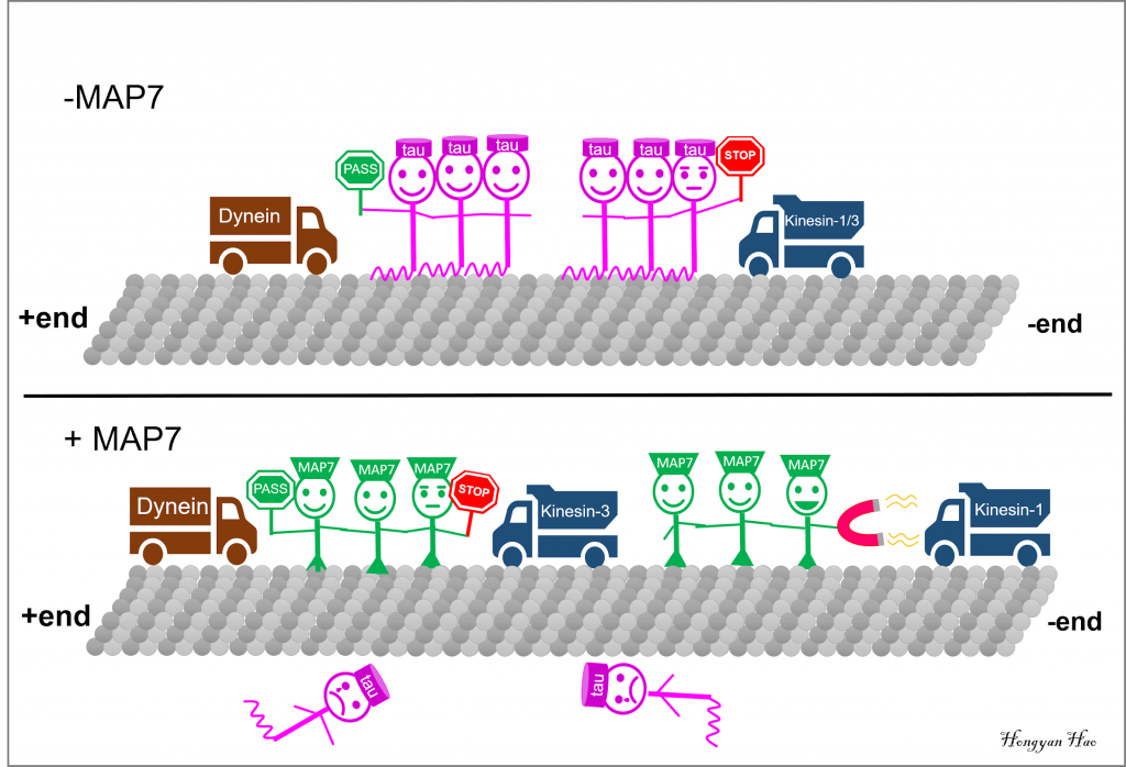
Microtubule (MT) plus-end motor kinesin-1 and kinesin-3 are inhibited by tau patches, while tau has little effect on the MT minus-end motor dynein. MAP7 outcompetes tau and specifically enhances kinesin-1 movement. Similar to tau, MAP7 inhibits kinesin-3 but allows for the dynein movement.

In axons, MTs are highly organized into bundles with plus ends directed out towards the cell periphery, and minus ends oriented inward towards the cell center. In contrast, MT orientation is mixed in dendrites (1). In vivo, MT organization and dynamics are highly regulated by microtubule associated proteins (MAPs). For example, the MAP tau binds to the MT lattice and stabilizes axon MTs. When detached from the MT, tau proteins form aggregates, which is one of the hallmarks of Alzheimer’s disease (2). Another MAP, MAP7, also known as ensconsin or EMAP-115, binds to MTs at axon branch sites to positively regulate branch formation (3). As the MTs are decorated by different kinds of MAPs in axons and dendrites, how do the motors dynein and kinesin behave when they hit MAPs?

MAPs are not just rocks on the road that block motors. In fact, the two motors will respond differently depending on which MAP they encounter. For instance, previous studies have shown that kinesin-1 motors will stop and fall off when they encounter tau, while tau has little effect on dynein (4, 5). In contrast, MAP7 enhances kinesin-1 movement (6, 7). How do these different motors and MAPs coordinate with each other to achieve effective cargo transport in axons and dendrites?

A recent publication by BMCDB student Brigette Monroy and her colleagues from Kassandra Ori-McKenny’s lab at UC Davis explores how motor movement might be regulated by different MAPs coating the MTs (8). Consistent with previous reports, Monroy et al. found that tau is restricted to axons, but MAP7 was localized to both axons and dendrites in the Drosophila peripheral nervous system and tissue cultured mouse neurons. Using Total Internal Reflection Fluorescence (TIRF) microscopy, which allows for visualization of molecules on a single stabilized MT in vitro, the authors found that MAP7 strongly excluded tau from the MTs. Although bound to MTs more rapidly than MAP7, tau is displaced by MAP7 over time.  This is most likely due to MAP7’s higher MT-binding affinity and longer dwell time on the MT lattice since purified MAP7 and tau do not bind to each other (8).

It is striking that MAP7 kicks tau off the MTs, but what are the physiological functions of MAP7/tau competition? Overexpression of MAP7 in Drosophila larval dendritic arborization (DA) neurons caused increased branch number while tau overexpression led to a decreased branch number. Overexpression of both MAP7 and tau led to more branches in DA neurons, consistent with the observation that MAP7 outcompetes tau (8).

One possible mechanism is that MAP7 and tau affect DA neuron branching by regulating the intracellular transport of the Golgi outposts. Golgi outposts are important for branch formation by nucleating MTs at branch sites (3, 9). The increased branching observed with MAP7 overexpression may be due to increased kinesin-1 mediated cargo transportation since Golgi were found to be enriched in the plus ends of MTs in DA neurons.

In vivo studies have revealed positive regulation of kinesin-1 by MAP7, but the mechanism has been unclear (7). Using in vitro single molecule motility assays, where the movement of a single fluorescently labeled motor protein on the MT is observed under TIRF microscopy, Monroy et al. found that both human and Drosophila MAP7 directly affect kinesin-1 by enhancing the motor landing on the MT. This may be due to 34 conserved amino acids in MAP7 which may facilitate kinesin-1 recruitment through a weak, ionic interaction with kinesin-1. Kinesin-1 motors stop and fall off the MTs when they encounter patches of tau. Would the presence of MAP7 overcome tau’s inhibition on kinesin-1? The answer is yes. MAP7 can not only replace tau on the MT and recruit kinesin-1 onto the MT, but also restores the ATPase activity of kinesin-1 inhibited by tau.

Tau has little effect on dynein motors. What about MAP7? Interestingly, just like tau, MAP7 barely affects the mobility of the dynein active complex in vitro. So, does MAP7 only affect plus end MT motors? Kinesin-3 family motors are also plus end directional. In contrast to kinesin-1, MAP7 inhibits kinesin-3’s landing on the MT as well as its mobility. However, tau inhibits kinesin-3 in a similar way as kinesin-1.
Cells have very complex environments and it is challenging for in vivo studies to provide clear and detailed mechanisms of how proteins work with one another. On the other hand, in vitro results can be difficult to interpret because simplified systems introduce biases not present in cells. In this paper, the Ori-McKenny lab illustrates how powerful it is to combine in vitro and in vivo studies to investigate the mechanism of cellular processes.

MTs are essential to maintain the shape, polarity and intercellular transport in our neurons. The findings suggest that MAPs on the MTs coordinate with each other and that MT motors can interpret MAPs, which allows for the regulation of cargo transport. Monroy’s story only discussed MAP7 and tau, but MTs are also occupied by other MAPs. Also, MTs can be post-translationally regulated to favor or inhibit motor mobility. I am grateful that our neurons have a system to coordinate cargo transport, which makes up a powerful neural system that enables me to ponder the relationship between different MAPs, motors and how they are doing their jobs together on the MTs! I am excited to see what Monroy and Ori-McKenney come up with next! A more complex map about motors and MAPs is yet to be revealed!

Now, after sitting here for a while, I have decided to walk out to get a cup of iced tea. I suppose that the kinesin and dynein motors made their ways to the right destination and the long motor neuron axons delivered the message to my toes, since I made it to the long line and am enjoying it now.


References:
1.         Burton PR (1988) Dendrites of mitral cell neurons contain microtubules of opposite polarity. Brain Res 473(1):107-115.
2.         Ballatore C, Lee VM, & Trojanowski JQ (2007) Tau-mediated neurodegeneration in Alzheimer’s disease and related disorders. Nat Rev Neurosci 8(9):663-672.
3.         Tymanskyj SR, Yang B, Falnikar A, Lepore AC, & Ma L (2017) MAP7 Regulates Axon Collateral Branch Development in Dorsal Root Ganglion Neurons. J Neurosci 37(6):1648-1661.
4.         Ebneth A, et al. (1998) Overexpression of tau protein inhibits kinesin-dependent trafficking of vesicles, mitochondria, and endoplasmic reticulum: Implications for Alzheimer’s disease. Journal of Cell Biology 143(3):777-794.
5.         Dixit R, Ross JL, Goldman YE, & Holzbaur EL (2008) Differential regulation of dynein and kinesin motor proteins by tau. Science 319(5866):1086-1089.
6.         Barlan K, Lu W, & Gelfand VI (2013) The Microtubule-Binding Protein Ensconsin Is an Essential Cofactor of Kinesin-1. Current Biology 23(4):317-322.
7.         Metzger T, et al. (2012) MAP and kinesin-dependent nuclear positioning is required for skeletal muscle function. Nature 484(7392):120-+.
8.         Monroy BY, et al. (2018) Competition between microtubule-associated proteins directs motor transport. Nat Commun 9.
9.         Ori-McKenney KM, Jan LY, & Jan YN (2012) Golgi Outposts Shape Dendrite Morphology by Functioning as Sites of Acentrosomal Microtubule Nucleation in Neurons. Neuron 76(5):921-930.

Say Cheese! A Snapshot of Microbial Communities on Cheese

Author: Jessica Huang

Editors: Keith Fraga, Hongyan Hao, Sharon Lee


I love eating cheese. If you set a block of cheese in front of me, I will probably not stop eating. One of my favorite memories from taking French in high school, besides singing a bunch of Disney songs in French, was the time my teacher brought a few different cheeses for us to try out. I have briefly thought about getting a cheese wedding cake if I ever manage to get married.

But what makes cheese so cheesy? The answer is, surprisingly, microbes.


How is cheese made?

Before we delve into the contribution of the microbial community within cheese, it’s important to know a bit about how cheese is made. The key component in milk for making cheese is the protein casein. Normally, casein is found in a micelle form in milk, which means that several caseins aggregate together into a spherical form. Since casein is negatively charged, the spherical micelles repulse one another. However, if acid is added, the negative charge is neutralized, allowing for coagulation, which is the thickening of milk into solid curds that are used to make cheese. Using this method alone will produce softer cheeses.

Comic about caseins coming together.


Another way to promote coagulation is to use rennin, which contains a protease called chymosin that breaks down κ-casein, a soluble protein that forms a protective and stabilizing layer around casein micelles. This formation of the solid milk curds is the first step in making cheese. The curds are then separated from the whey, which is a liquid byproduct composed of proteins that remain soluble even after acidification and rennet treatment. Whey can be used for several things, such as protein shakes. Meanwhile, the curds are processed in various ways to make different types of cheeses. The last step is the maturation of cheese, which contributes most to the distinct flavor of cheese and can last anywhere from several months to several years.


So where do microbes come into the picture?

While vinegar or lemon juice can be used to acidify milk, another common method is to use microbial starter cultures instead. Some of the most commonly used strains in starter cultures include Lactococcus lactis (used for cheddar), Streptococcus thermophilus (used for mozzarella), and the Lactobacillus species (used for Swiss cheese). These bacteria help acidify milk by converting lactate into lactic acid.

Microbes also play a significant role during the aging process of the cheese. During this time, bacteria added from the starter cultures begin to die off, while other microbes that were either already in the milk from the beginning or joined in from the environment along the way begin to flourish.

Sometimes additional microbes known as adjunct cultures are added. Their main purpose is to enhance the flavor or texture of the cheese rather than to produce lactic acid. One example is the mold Penicillium roqueforti, which puts the blue in blue cheese through its formation of spores. The unique flavor of blue cheese comes from the conversion of fats into flavorful free fatty acids and methyl ketones by lipases produced by P. roqueforti. Another example is Penicillium camemberti, which gives cheeses like camembert and brie their soft, gooey texture. Its preference for the surface of the cheese allows for the development of a characteristic soft, white, bloomy rind. The holey appearance of Swiss chese is thanks to Propionibacterium freudenreichii. These bacteria convert lactic acid inside the cheese into carbon dioxide, which becomes trapped and forms bubbles. Acetate and propionic acid are also produced as byproducts and contribute to the flavor of the cheese. P. freudenreichii is often used together with Lactobacillus helveticus, a strain that can also be used when making cheeses like cheddar and that helps produce a nutty flavor.


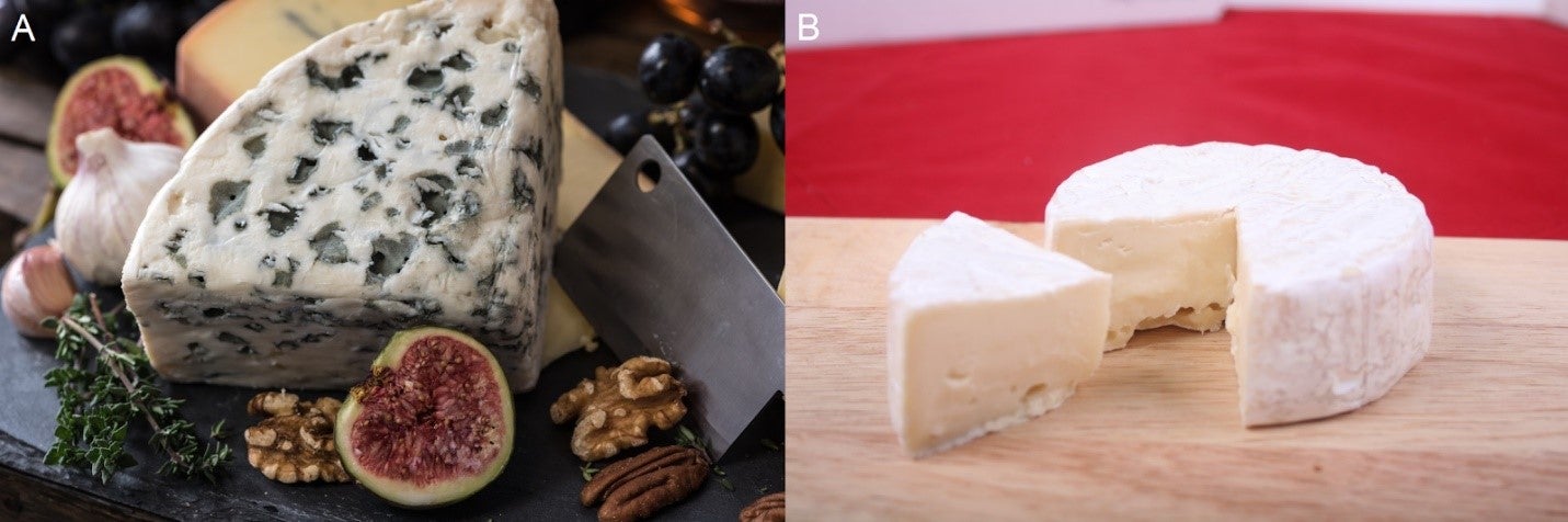
A) Spores formed by  roqueforti throughout blue cheese. B) Bloomy rind of brie formed by P. camemberti.


Of course, there are many other microbes present in cheeses. In addition to those that are added, there are also microbes that were present from the very beginning in the milk, as well as ones that were acquired throughout the cheese-making process. Furthermore, many factors such as pH, temperature, and salt content affect which microbes are capable of forming colonies in the cheese. The presence of the microbial community, the metabolic byproducts they produce, and their interactions with one another in turn produce the distinctive flavor, aroma, and texture of many different cheeses.


A cheesy model system?

Cheese is home to many different microbes, and the rind is especially rich in microbial diversity. This makes cheese a potential model system for studying microbial communities. One of the labs that uses cheese for research is headed by Rachel Dutton at UC San Diego. Back in 2014, her lab successfully sequenced 136 different rind communities and found that they are highly reproducible. They discovered that the composition of the microbial community on cheese rinds and the relative abundance of individual microbes is correlated with the type of rind. For example, the bloomy rinds of camembert have a denser population of fungi, as would be expected of rinds produced due to the mold P. camemberti. The environment that the cheese is exposed to as it ripens is important too, but surprisingly, where the cheese was made does not correlate with the composition of the microbial community.

The Dutton group also found that many of the dominant species can be isolated and cultured, allowing them to easily recreate the communities in vitro. Thus, they were able to study interactions between different species, and observe how they affect one another in pairs. One of their findings is that the presence of fungi may promote an increase in pH, creating an environment that is beneficial to some bacteria. Overall, it looks like they’ve succeeded in creating quite the tasteful system.


Cheese is grate

Cheesy pun aside, cheese really is quite wonderful. It’s the basis of many delicious dishes and makes for a great snack by itself. On top of all that, it’s also making cool contributions to science. You gouda love it.

(P.S. Cheese can still go bad. If you see mold that isn’t supposed to be on the cheese, throw it out if it’s a soft cheese. For hard cheeses, which have lower moisture content, you should be able to cut off the moldy part and keep the rest. Bon appetit!)


References

  • Button JE, Dutton RJ. Cheese microbes, Current Biology 22 (2012) R587-R589.
  • Wolfe BE et al. Cheese Rind Communities Provide Tractable Systems for In Situ and In Vitro Studies of Microbial Diversity. 158 (2014) 422-433.
  • https://www.cheesescience.org/microbes.html#lab


Not so devious intentions

When people are doing annoying things that affect you, how often do you feel they were intentionally direct towards you? Do you feel rage when someone cuts you off on the highway because you are sure they are just a**holes trying to take advantage of you? Do you think people are trying to sabotage your experiment when moving your -80C box from one location to the other? Well, sometimes yes, but often there is an alternative explanation. Someone could just be trying to rush to the hospital or simply misplaced your freezer box when searching through the racks during a rushed experiment. We often fixate on the potential negative intentions when there are plenty other possible explanations. Considering alternatives will lead you to reduce your bad days and avoid some unnecessary grudges. Hopefully, this short YouTube video from the School of Life can help you develop the skills to not rush-to-judgment on the benign intentions of others.  


-Yulong


This post is edited by Keith Fraga

For any content suggestions or general recommendations, please email to UCDBioScope@gmail.com and put science 2.0 in the title

Difficult Conversations

It’s often difficult to talk about sensitive topics that involve strongly held personal beliefs.  This is especially true when that person is close to you and you don’t want to damage your relationship. Sometimes those conversations can be avoided, but do you have a plan on how to strike those conversations if they are too important to be avoided? As a scientist, I often feel obligated to promote science, particularly to the most skeptical crowds. For example, contending with the views anti-vaxers. This podcast from The New York Time’s Change Agent has some helpful insights on how to have those conversations. Specifically, the strategies (validation, getting curious, and personal stories) developed from an ex-cult member turned mental health counselor Steven Alan Hassan.


-Yulong


https://www.stitcher.com/podcast/the-new-york-times/change-agent-2/e/53526869


This post is edited by Keith Fraga

For any content suggestions or general recommendations, please email to UCDBioScope@gmail.com and put science 2.0 in the title.

Mentoring and being mentored, the Graduate School Edition

Author: Sharon Lee

Edited by: Keith Fraga


I remember the day I walked into her office for an interview.

Dr. Tama Hasson, Director of the Undergraduate Research Center-Sciences at UCLA, had been doing this for a while and knew that a sure way to comfort a nervous student was her big, encouraging smile. Before I knew it, within the next fifteen minutes of our meeting, she laid out my entire academic plan and became one of my first mentors! At that time, I only knew about my interest in research. I had just joined a lab to satisfy my growing scientific curiosity. But I didn’t know anything about graduate school.

As a first-generation college student and the only one in my family pursuing a PhD, I am grateful for the support and guidance I received through the years from my mentors! Finding good mentors has been a skill I have tried to develop over the years and I hope to share a few things I have picked up about how to be mentored.


What is mentoring and how important is it to find a good mentor?

Image credit: “Piled Higher and Deeper” by Jorge Cham www.phdcomics.com

I like the definition of mentoring from the American Association of Pharmaceutical Scientists: mentoring is a relationship between two individuals (a mentor and a mentee) based on a mutual desire for development towards career goals and objectives.

In graduate school, the most important mentoring relationship is the one that develops between graduate students and their thesis advisor (research mentor). It is also one of the most sensitive relationship and sometimes, can be challenging to maintain.

As an undergraduate, all I focused on was the type of research I was interested in. When I was looking into labs to join, I prioritized the research topic over everything. It was not until much later that I realized finding a good faculty mentor is more important than working on a particular project. First-year graduate students tend to forget this when choosing their thesis labs.

Dr. Daniel Starr, Professor and former BMCDB Chair, agrees that a match between a graduate student and a faculty mentor is more important than the project. As scientists, we should be excited about a variety of different projects, but “the match is essential” for a successful and nourishing graduate career.

To collect some thoughts about mentoring from other faculty and students, I circulated an anonymous mentoring survey within the BMCDB graduate group. All 18 faculty who responded to the survey also agreed that good mentoring is very important for a graduate student’s training.


What is key for a successful mentoring relationship?

When asked what is key for a successful mentoring relationship, the response that I received from the majority of faculty and students was communication. Clearly communicating one’s expectations from a mentoring relationship helps to avoid any misunderstandings that may arise from unspoken assumptions about the roles and responsibilities of a mentor and mentee.

Dr. Starr, who has mentored 9 graduate students, also added that because “every student needs a different mentoring style”, mentors need to be “flexible and patient” with their students. It was encouraging to see that 94% of the BMCDB faculty surveyed were open to changing their mentoring style to meet the needs of their graduate students.

In addition to communication, Dr. Steve Lee, Graduate Diversity Officer from UC Davis Graduate Studies, believes an important factor for a successful mentoring relationship is self-awareness. Self-awareness is critical because both mentors and mentees need to recognize the way they best communicate their ideas and how they best receive feedback. A high level of self-awareness helps recognize when there is a dissonance between you and your mentor/mentee. It allows you to put aside your differences and work collaboratively to meet shared goals.

Dr. Lee suggests that regular self-assessment is a good practice for graduate students to understand where they stand. Particularly for women and students from minority backgrounds who experience higher levels of imposter syndrome, communicating with one’s mentor can be intimidating. Self-assessment can be used as a strategy to overcome imposter feelings and take actions to move forward. Dr. Lee recommends for the students to mentor up!


What is Mentoring up?

“Mentoring up is a concept that empowers mentees to be active participants in their mentoring relationships by shifting the emphasis from the mentors’ responsibilities in the mentor-mentee relationship to equal emphasis on the mentees’ contributions.

This term was conceptualized by Dr. Lee with colleagues back when he served as the Assistant Director of CLIMB. They came up with this idea to encourage students to proactively engage with their research mentors for an effective mentoring relationship.

The core principles and framework of mentoring up is described in Chapter 7: Mentoring Up”: Learning to Manage Your Mentoring Relationships, in the book, “The Mentoring Continuum – From Graduate School through Tenure”. In this chapter, the authors provide strategies for mentees to consciously contribute and guide their mentoring relationships through difficult situations, avoiding passive patterns of behaviors that may limit their own success.

I highlighted below three of the seven core principles and strategies of mentoring up that graduate students (mentees) can use to foster their mentoring relationship:

  1. Maintaining Effective Communication. It is important that mentors and mentees seek to understand each other’s communication styles and take time to practice communication skills, particularly when their preferred method of communication is different.
    • Determine your mentor’s preferred medium of communication and acknowledge if it differs from your own personal preference.
    • Schedule a regular time to meet with your mentor and prepare for the meetings by articulating specifically what you want to get out of the meeting.
    • Keep track and share progress toward project and professional goals, both verbally and in writing.
  2. Aligning Expectations. With clear expectations, mentoring relationships are more likely to be productive. Problems and disappointment often arise from misunderstandings about expectations. In order to avoid such misunderstandings, expectations should be clearly discussed and realigned on a regular basis as they may change over time.
    • Ask your mentor for his or her expectations, and also share yours, regarding your research project, role and responsibilities of being a graduate student, and your professional career goals.
    • Ask others in your lab about your mentor’s explicit and implicit expectations.
    • Write down the expectations you agree to with your mentor and revisit them often.
  3. Assessing Understanding. Determining how well you understand your mentor as well as how well your mentor understands you is not easy, but is crucial for a productive mentoring relationship. Develop strategies to critically assess each other’s understanding.
    • Take a minute to consider any assumptions you have made about what your mentor knows or does not know about how well you understand your project.
    • Ask questions when you do not understand something. If you are afraid to ask your mentor directly, start by asking other lab members and peers.
    • Explain your project to someone who is not familiar with your field and help them to understand your project and its significance.

I asked my thesis advisor once to re-explain some concepts about my research project which I did not clearly understand. I was worried that he would be unhappy about having to repeat himself. But in hindsight, I am glad that I asked my questions because my advisor was equally pleased to have clarified my confusion. He actually appreciated my initiative to clear my misunderstanding.

We students so often assume that our advisor will react negatively to our mistakes and lack of understanding, which leads us to make an even bigger mistake of not openly communicating. 75% of the students surveyed reported that despite being not extremely happy with their thesis advisors, they did not talk with them about improving their relationships.  

Image credit: “Piled Higher and Deeper” by Jorge Cham www.phdcomics.com


How to get the best of all worlds?

One of the common mistakes that graduate students make during their early years is to expect their thesis advisor to fulfil all the different roles and responsibilities of a mentor. They often need to be reminded that no one can do it all, that one mentor cannot provide all the guidance and support that a student needs. Effective mentoring is a community effort and thesis advisors should encourage and assist their students with finding other mentors with complementary skills and knowledge. Some examples of people who can make an excellent additional mentor include senior PhD students, postdocs in your lab, faculty you rotated with, and your thesis committee members. It is also a good practice to maintain your relationship with old mentors from previous institutions.  

Graduate students can also actively seek out mentors on their own. As a woman in STEM, I look for opportunities to engage with female scientists and identify women mentors. I love this quote from The Atlantic about How Women Mentors Make a Difference in Engineering: “It’s not that having a female mentor increased belonging or confidence – it just preserved it.” “They act as a “social vaccine” that protects female students against negative stereotypes and gives them a sense of belonging.”

Do remember that building an effective mentorship takes time and maintaining it takes effort. Mentorship is a work-in-progress and a long-term investment! Practice humility and be willing to adjust or compromise to achieve your mentorship goals. However, if the relationship is not working, take charge and do something about it. I greatly appreciated when Dr. Starr encouraged students to do the same. He said that “students need to know that some matches don’t work, like a large percentage of matches don’t work” and it is fine to change labs and find a new mentor who can help you be successful. “Graduate school is hard enough as is … hopefully you find a match that makes graduate school fun, hard but fun.”


Resources:

  • National Research Mentoring Network (NRMN): NRMM is a nationwide consortium of biomedical professionals and institutions collaborating to provide trainees across the biomedical, behavioral, clinical and social sciences with evidence-based mentorship and professional development programming.
  • Questionnaire for Aligning Expectations in Research Mentoring Relationships: a tool to help beginning graduate students to align their expectations with their thesis advisors. Also helpful in assessing where you stand compared to other graduate students in the lab, among your cohort, and/or within the graduate group.

Page 2 of 3

Powered by WordPress & Theme by Anders Norén