A UC Davis Graduate Student Blog

Category: main_pub Page 2 of 4

The Science Behind Going Keto

Written By: Ross Wohlgemuth

Edited By: Sydney Wyatt


In the plethora of dieting options out there, it can be difficult to find the right one for you; sticking with it and seeing the results weeks or months later can be even more challenging. Whether it’s a traditional low-fat diet (LFD) or something more complex like intermittent fasting, each eating regime has its pros and cons that apply to different types of people with different goals in mind. Often, people choose the wrong diet or fail to stick with it, and do not reach their perceived goals. In worse cases, people may develop or worsen eating disorders by following strict diets. There is also a lot of misinformation about dieting, so it would be a shame if your 2020 resolution centered around a diet that has no scientific backing. Whatever the case may be, finding the right diet and sticking with it should be done with medical supervision.

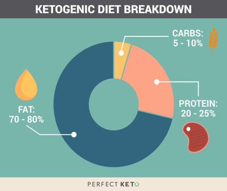
Fig. 1. Sample ketogenic diet macronutrient proportions. This differs from the USDA recommendation by a large amount, especially in the carbohydrate category. Image from [3].

One diet in particular that is popular for weight loss and athletic performance is the ketogenic diet (KD, keto). The KD is similar to many diets in that it cuts carbs, almost to zero. While the USDA recommends that 45-60% of calories come from carbohydrates [1], the KD suggests less than 50g [2]. The macronutrient deficit is made up by an increase in fats, which make up around 70% of the caloric intake on keto [2].

The basis of the KD is that when your carbohydrate intake is low, your body can adapt by producing a new energy source called ketone bodies (KBs) [4]. KBs are made by mitochondria in the liver from fatty acid derived acetyl-CoA, and circulate throughout the body and even cross the blood-brain barrier to provide a source of energy to your cells [4,5] (Figure 2). Thus, even without consuming the recommended amount of carbohydrates, your body can still function just fine by running on fats—for the most part. The jury isn’t out on keto just yet, as some doctors and health organizations are still hesitant to proclaim as a safe way to eat.

For most people, one of the biggest factors in choosing a diet is how effective it is at promoting weight loss. In the case of the KD, science seems to support its weight loss promotion. In a meta-analysis [6] on thirteen studies involving long-term weight loss on a KD or a LFD, it found that five of the parameters tested differed between the KD and LFD groups, one of them being weight loss. They found that the average weight loss of the KD groups was 2 pounds more than that of the LFD groups. The study also found KD groups experienced decreases in blood triglycerides and diastolic blood pressure and increases in both high-density and low-density cholesterol (HDL, LDL).

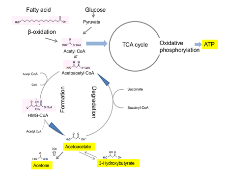
Fig. 2. Schematic of ketone body (KB) formation. 3-Hydroxybutyrate, a KB, is produced from the oxidation of fatty acids, resulting in the production of acetyl CoA. Acetyl CoA is consequently converted into HMG-CoA and then to acetoacetate which is the precursor to 3-hydroxybutyrate. Figure and caption adapted from [5].

At first glance, this seems like a solid win for the KD since 3 out of the 5 changes are considered to represent indicators of health, but there are limitations to these studies to consider. The studies took place over a year or longer, and the provided counseling from a dietician varied between studies. In addition, participants were obese individuals with a mean age between 40 and 60. Further, the amount of carbohydrate intake per day by each participant was only reported to be in the keto range of 50g or less in one of the studies; the other studies either had a greater caloric proportion allocated to carbohydrates, or the value was not reported. Even greater concern is the high participant drop-out rate (15-85%) from the studies. This variation makes it hard to trust these studies, and the KD regime in general. Ultimately, the 2 pound difference between the keto and low-fat diets over a year’s time is not that much of a change, even if it is statistically significant. Thus, even though this study attempts to demonstrate that the KD is more effective in producing weight loss than the traditional LFD, there is still much to question as to the ability of participants to stay on the diet, and the relevancy of the results to a younger or more fit population.

Another factor to consider when choosing a diet is whether it improves your physical fitness or athletic performance. Under the scrutiny of scientific investigation, it seems that the KD may only benefit athleticism in certain cases. In a pilot study of five endurance athletes (four female and one male) between the ages of 49-55, a KD was maintained over 10 weeks of each athlete’s typical training schedule, consisting of 6-12 hours per week of cycling or running [7]. The athletes were tested for body composition and athletic ability at the end of the period, and were also asked to give commentary about the experiences they felt during their time on the diet.

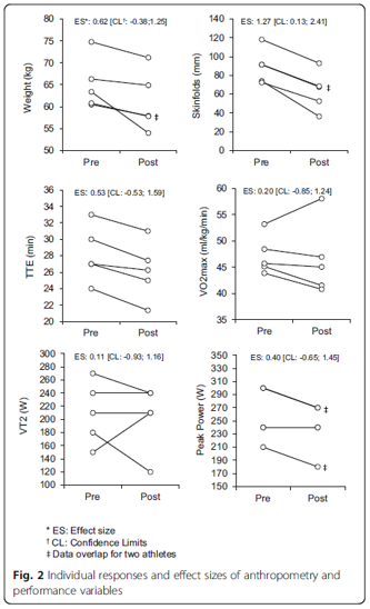
Fig. 3. Weight loss, time to exhaustion (TTE), and ventilatory threshold (VT2) in endurance athletes from [6].

The researchers found that the body composition of the athletes improved significantly, with an average weight loss of almost 9 pounds and a significant loss in fat mass as measured through several skin folds (Figure 3). This positive weight loss was not accompanied by positive results in performance however, with declines in VO2 Max, peak power, and ventilatory threshold (Figure 3). Some athletes even reported negative experiences toward the beginning of the study, including complaints of fatigue or irritability, however, there were more positive experiences reported like feeling healthier or enjoying the weight loss by the end.

The researchers concluded that although the KD was effective in improving body composition and weight, it was not successful in improving endurance performance. They hypothesized that the lack of athletic improvement was due to lower rates of glycogenic metabolism (metabolism which uses glucose as the primary substrate), which could hinder performance in high intensity exercise. This could be due to lower blood insulin downregulating pyruvate dehydrogenase, an enzyme necessary in linking the glycolytic pathway in the cytosol to the Krebs cycle in the mitochondria [8]. The researchers also thought that liver glycogenolysis (breakdown of glycogen) could be influenced by dietary intake of carbs while gluconeogenesis (the process of making new glucose from lactate, glycerol, and other substrates) remained constant during KD [9]. Whatever the case may be, it seems that using KBs as a metabolic substrate attenuates the athletic ability of the endurance athletes in maximal intensity efforts.

In another study that compared the effects of the KD and traditional western diet (WD) on strength performance in nine elite male gymnasts around the age of 21 [10], researchers found that 30 days of the KD produced better changes in body composition than the WD, but not in strength or performance. Since the athlete’s training regimes were consistent throughout the year, each participant was able to undergo 30 days of the KD and 30 days of the WD in order to pair the two treatments for comparison (a 3 month gap was used between the two dieting periods). The results showed that 30 days of the KD led to an average weight loss of 3.5 lbs and a fat mass reduction of 4.2 lbs. This translates to a change in body fat percentage of 2.6% and an increase in lean mass percentage by a similar degree. Nevertheless, these underlying changes in body composition did not affect the athlete’s muscle mass or their athletic ability. There were no significant changes in either parameters after the KD or WD regimes. The investigators attributed the weight loss to fullness from adequate protein consumption, a greater ratio of fat breakdown to fat synthesis, lowered resting respiratory exchange ratio (meaning the predominant fuel during rest was fat), and elevated metabolism from gluconeogenesis and the thermic effect of proteins [11]. They also stated that increasing muscle mass during the KD is difficult since blood insulin levels are so low, which attenuates the muscle growth pathway via IGF-1, mTOR, and AKT (Figure 4) [12]. Thus, maintaining muscle mass during the KD is a more reasonable and attainable goal than gaining mass. With that in mind, the relevance of the KD to athletic performance becomes more important to athletes whose sport involves weight classes such as boxing. Competitors who desire to maintain muscle mass and strength while trying to lose fat to fit into a lower weight class may reasonably benefit from short term use of the KD. With this in mind, the KD seems like a reasonable option for a specific niche of athletes who may stand to gain (or lose) from periodic keto use.

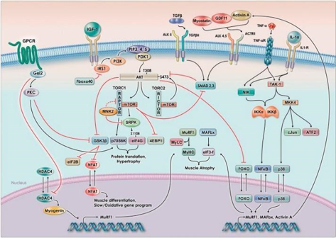
Fig. 4. The signaling pathway for muscle hypertrophy relies on insulin and IGF-1 signaling, which leads to the increase in phosphorylation of AKT and the activation of mTOR. mTOR activation leads to downstream effects ultimately resulting in muscle hypertrophy (muscle growth). Figure taken and caption adapted from [12].

After looking at the KD as a weight loss promoter and performance enhancer, it certainly has its fair share of pros and cons. As far as diet commitment, the KD seems to be difficult in the first few weeks but easier as it continues. Not only were there great proportions of participants who dropped out from the studies involved in the meta-analysis, but the experiences reported from the endurance study show that the KD can be difficult and perhaps even miserable in the beginning. In addition, the carbohydrate caloric contents of the studies in the meta-analysis were not even low enough to be considered “keto” by the latter two studies addressed [7,10], so adherence to the KD by non-athletes or those who have less dietary or physical motivation may be called into question.

In the studies on endurance and strength athletes, the net carbs consumed per day were well below the 50g mark, with around 10-35g in the endurance study and 22g in the strength one. These bode well with the body composition and weight loss observed in the short amount of time the KD was followed compared to the long-term studies in the meta-analysis. Even though the meta-analysis showed that the KD had better weight loss effects than the LFD, the amount of weight lost in the 12-month period was unimpressive compared to the short-term studies. This may have to do with the demographics of each population, as the long-term studies were done on older, obese individuals and the short ones done on active people of various ages. This highlights the confounding effect of lifestyle and level of exercise to dietary studies of the KD. It is virtually undeniable that diet and exercise are the cornerstones of weight loss programs, and the combination is more effective than either alone.

The KD presents some promise to those looking to lose weight who are also generally active on a weekly basis. Although it may not be useful in improving athletic performance, it is remarkably beneficial to metabolic health when used safely and correctly. Again, any new diet program you want to try should be discussed thoroughly with a qualified physician or dietician. Given the scientific literature on the KD, I would say KD shows promise for a certain niche of people who are focused on losing weight.

Citations

  1. Dietary Guidelines for Americans. 2010. USDA, USDHHS.
  2. Gunnars, Kris. (04 January 2019). 5 Most Common Low-Carb Mistakes (And How to Avoid Them). Healthline.com 
  3. Ketogenic diet breakdown. Perfectketo.com
  4. Pinckaers, P.J.M., Churchward-Venne, T.A., Bailey, D., van Loon, L.J.C. (2017) Ketone Bodies and Exercise Performance: The Next Magic Bullet or Merely Hype? Sports Med. 47:383-391. doi:10.1007/s40279-016-0577-y
  5. Watanabe, Shaw & Hirakawa, Azusa & Aoe, Seiichiro & Fukuda, Kazunori & Muneta, Tetsuo. (2016). Basic Ketone Engine and Booster Glucose Engine for Energy Production. Diabetes Research – Open Journal. 2. 14-23. 10.17140/DROJ-2-125.
  6. Bueno, Nassib Bezerra, et al. “Very-Low-Carbohydrate Ketogenic Diet v. Low-Fat Diet for Long-Term Weight Loss: a Meta-Analysis of Randomised Controlled Trials.” British Journal of Nutrition, vol. 110, no. 7, 2013, pp. 1178–1187., doi:10.1017/S0007114513000548.
  7. Zinn, C., Wood, M., Williden, M., Chatterton, S., Maunder, E. (2017) Ketogenic diet benefits body composition and well-being but not performance in a pilot case study of New Zealand endurance athletes. Journal of the International Society of Sports Nutrition. 14(22) doi:10.1186/s12970-017-0180-0
  8. Peters, S.J., LeBlanc, P.J. (2004) Metabolic aspects of low carbohydrate diets and exercise. Nutrition and Metabolism. 1(1):7. doi: 10.1186/1743-7075-1-7.
  9. Webster, C.C., et. al. (2016) Gluconeogenesis during endurance exercise in cyclists habituated to a long-term low carbohydrate high-fat diet. The Journal of Physiology. 594(15): 4389-4405. doi:10.1113/JP271934.
  10. Paoli, A., Grimaldi, K., D’Agostino, D., Cenci, L., Moro, T., Bianco, A., Palma, A. (2012) Ketogenic diet does not affect strength performance in elite artistic gymnasts. Journal of the Society of Sports Nutrition. 9(34)
  11. Paoli A., Canato M., Toniolo L., Bargossi A.M., Neri M., Mediati M., Alesso D., Sanna G., Grimaldi K.A., Fazzari A.L., Bianco A. (2011) The ketogenic diet: an underappreciated therapeutic option? La Clinica Terapeutica. 162:e145–e153.
  12.  Egerman, M.A., Glass, D.J. (2014) Signaling pathways controlling skeletal muscle mass. Crit Rev Biochem Mol Biol. 49(1): 59-68. doi:10.3109/10409238.2013.857291


Working Distractions: the cost and ways to overcome  

Written By: Ellen Osborn

Edited By: Emily Cartwright and Anna Feitzinger


At any given moment, there are multiple, maybe even dozens, of things that demand our attention as graduate students. It could be the unread emails that need to be responded to, the experiment that needs to be planned, the papers sitting at home that need to be graded, the grant that needs to be written, and the list goes on. Whether it is an effect of our world moving faster, or a consequence of ourselves growing older, it is becoming increasingly more difficult to focus on a single task and ignore incessant distractions. 


According to the 2018 Workplace Distraction Report published by the online learning company Udemy, Millennials and Gen Z were found to be the most distracted generations; 74% of surveyed individuals described themselves as distracted at work. Millennials and Gen Z are distracted at school as well as work: a cohort of university students were found to focus on a single task for only 6 minutes before succumbing to a distraction (Rosen et al., 2013). 


While this might not come as a surprise, our brains do not do their best work when we are distracted. Business professor Sophie Leroy coined the term attention residue, defined as the attention that remains with an initial task even when an individual has transitioned to a second task. In experiments that involved giving participants a cognitively demanding task to complete, such as solving a complex puzzle, it was found that for those participants that were briefly distracted, even by just glancing up at a picture, their performance dropped significantly when returning to the original task. Just by changing their context very briefly, the attention residue seized by the distraction prevented individuals from performing at their best, and not just momentarily. It took some time before the attention residue fully cleared and individuals were again fully focused on the task at hand. Dr. Leroy’s work echoes the findings from a University of California, Irvine study where researchers shadowed individuals while at work and found that it took an average of 23 minutes for those individuals to get back to an initial task after being abruptly interrupted by either a telephone call or text message.


Cal Newport, professor and author of the cult book “Deep Work: Rules for Focused Success In a Distracted World,” makes the conclusion that by changing our cognitive context frequently at work, we are consistently building up attention residues that prevent us from ever truly focusing on a single task. That is, every time we take a quick glance at our inbox or attempt to multitask, we do so at the cost of performance. And while some can recognize this sacrifice of performance for distraction and address it with a casual “Well I guess I should be less distracted”, for those that rely on their brains to make a living (like graduate students), there should be more urgent concern. 


So what are some ways we can set aside everyday distractions in favor of developing more productive work habits? Pete Leibman, creator of StrongerHabits.com and bestselling author, promotes three basic steps to minimize attention residue throughout the day. First, instead of jumping from task to task rapidly during the day, try to focus on a single project per day. Or, if that is not possible, dedicate the morning to work on one project, and then focus on a second project in the afternoon. This removes the largest cause of attention residue: multitasking between several unrelated projects. Second, because it can be overwhelming and demotivating to try to tackle a whole project in a single day, try breaking the project down into unambiguous pieces that can be completed in approximately 60 minutes. This practice will minimize the attention residue associated with working on related tasks at the same time, which is a sneaky form of multitasking. Third, plan to take short deliberate breaks throughout the day, especially when transitioning between tasks. Just like cleaning a paint brush before dipping into a new color, being intentional about taking short breaks in between tasks removes much of the attention residue that is stuck on the previous task. 


Some other practical tips offered up by professionals in the business of decreasing distractions: for one day, write down everything you do, any task, both major and minor (from checking Facebook to giving lab meeting). For each item you listed, ask yourself if that task is a distraction: something that kept you from focusing on the most important tasks in your day. If it is, intentionally plan your day so that you are only doing those distraction tasks during a defined block of time so they do not interfere with your more important tasks. Also, to prevent being caught wondering what task you should be focused on, plan your day the afternoon before (not waiting until the end of the day when you are less likely to do it). And one final tip, which may be the simplest but the most difficult: while at work, keep your phone silenced and tucked away where it cannot be seen or reached, eliminating the temptation to indulge in easy distractions. 


Related media to check out: this TedTalk for more on strategies to manage addictive distractions, and this NPR podcast with Cal Newport on deep work. 


Sources (in order of appearance): 

Udemy for Business. (2018). 2018 Workplace Distraction Report

Rosen, Larry D., L. Mark Carrier, and Nancy A. Cheever. “Facebook and texting made me do it: Media-induced task-switching while studying.” Computers in Human Behavior 29.3 (2013): 948-958.

Leroy, Sophie. “Why is it so hard to do my work? The challenge of attention residue when switching between work tasks.” Organizational Behavior and Human Decision Processes 109.2 (2009): 168-181.

Mark, Gloria, Daniela Gudith, and Ulrich Klocke. “The cost of interrupted work: more speed and stress.” Proceedings of the SIGCHI conference on Human Factors in Computing Systems. ACM, 2008.

“You 2.0: Deep Work.” Hidden Brain, NPR, 27 Aug. 2019, https://www.npr.org/transcripts/754336716.

Leibman, Pete. “Attention Residue: The Costly Side Effect of Switching Tasks.” StrongerHabits.com, 21 Mar. 2019, https://strongerhabits.com/attention-residue/.

Science communication for the middle ground

Written By: Will Louie

Edited By: Nina Sibonae Cueva

It is flu season, so I hope you have all gotten your flu shots! Vaccinations are arguably one of the most game-changing medical achievements of human civilization. Developed countries enjoy the eradication and suppression of some of the deadliest viral and bacterial diseases. However, since the publication of the original study fraudulently linking the Mumps, Measles, Rubella (MMR) vaccine to autism, the anti-vax movement is on the rise in the United States. And while scientists focus on the black-and-white of whether to vaccinate when debunking this claim, vaccine-hesitant people are still left in the middle. This group does not flat out reject vaccines, but are either fearful about the side effects, considering alternative vaccination schedules, or just distrustful of medicine in general. It is critical that we as scientists empathize and address the sources of hesitancy, a phenomenon that describes reluctance but not complete opposition to vaccination.


Many infectious diseases that terrorized our ancestors are but a memory thanks to mass vaccination. According to the Center for Disease Control and Prevention (CDC), the incidence of many infectious diseases have dropped by over 90% since the implementation of quality-controlled vaccines. Globally eradicating smallpox in 1980 was only possible through mass vaccination. Famously, Jonas Salk’s first successful polio vaccine in 1955 was immensely successful in pioneering mass vaccination, as millions of American families volunteered their children in Salk’s government-funded vaccine trials. Parents truly felt they contributed to a greater good, and the rapid transition from fears of losing their children to the disease within a single afternoon to near eradication of polio incidence highlighted an immense payoff in trust of a government program. Following worldwide administration, polio has been nearly eradicated, with sporadic transmission confined to inaccessible regions of Pakistan and Afghanistan. Despite these success stories, vaccine hesitancy has persisted, and understanding the perspective of those who are vaccine hesitant is imperative to improving our communication with the public.


Fears about autism


The most resilient debunked argument against vaccinating children is the fear that vaccines cause autism. Although the study was retracted, the damage was already done: this argument lingers among social media groups and anti-vax blogs. More worrisome is not that people still believe this unsupported causal relationship, but that anti-vaxxers prioritize preventing autism over preventing potentially deadly diseases. The fear is unfounded and counters collective efforts to destigmatize physical and mental disabilities. While the universal consensus among scientists is that no causation between vaccines and autism exists, it is uncertain whether this rebuttal alone is sufficient. Many vaccine-hesitant parents are still unsure whether this autism link is truly debunked, thanks to the mass amounts of information and disinformation circulating on the internet. We should also delineate the differences in health outcomes between autism spectrum and deadly infectious diseases. Even if this were true, the benefits outweigh the risks, and marginalizing those who are actually on the spectrum is not helpful. As scientists, we must improve our communication to clearly share the benefits and real risks of vaccination without alienating an already marginalized population. Shifting our focus from sole denial of MMR-autism causation to an emphasis that the benefits of MMR protection are worth the risks of side effects, addresses parental concerns for their children’s health in a non-judgemental manner.


Fears about government ethics and transparency


We all have a favorite conspiracy theory; I love the claim that “pigeons are not birds but actually government surveillance drones implemented to spy on urban civilian life” (citation not needed). Likewise, there is no shortage of conspiracy theories regarding vaccines. Moreover, there is a striking overlap between people who believe in conspiracy theories and those who are skeptical of vaccines. However, one cannot help being sympathetic to groups who are distrustful of government. After all, the U.S. government’s track record for medical ethics and transparency has been less than stellar. From the Tuskegee syphilis experiments targeting African American males on the domestic front to the CIA’s fake vaccination program in Pakistan as a cover for hunting Osama bin Laden on the international front, it is no surprise that many people are skeptical of government-mandated vaccine compliance. While there is still a giant chasm between outlandish chemtrail conspiracies and a real concern over government regulation or deregulation of vaccine administration, we need better ways to communicate the differences. Those who are vaccine-hesitant often struggle to separate distrust in the business practices of our corporate overlords from distrust in the science itself. Public health professionals and medical researchers need to bolster efforts at communicating the risks, benefits, and evidence for vaccines in a transparent and assuring manner. Most importantly, however, is the need to repair trust between the government and the people, a problem that transcends the anti-vax movement.


Fears about Big Pharma


Similar to the distrust in government is the distrust in companies that profit from vaccine development and distribution. It is easy to paint the pharmaceutical industry as the villain, because it is true in many cases. Examples of corporate greed taking priority over civilian health are all too numerous: former Turing CEO Mark Shkreli’s 5000% markup on the drug Daraprim (arguably a catalyst for discovery of the even more sinister predatory price gouging by Valeant Pharmaceuticals); Purdue Pharma’s role in fueling the opioid crisis; and Bayer knowingly selling HIV-tainted products to developing countries. As a self-proclaimed jaded millennial, I am not surprised that there are people, paranoid or misinformed, who see vaccines as just another case of Big Pharma capitalizing on a medical necessity to maximize profits. But Big Pharma generally doesn’t profit from vaccines. The cost of an annual flu shot ranges from $0 to $50 depending on your medical provider, while other vaccines like the intravenous polio vaccine are being given to children in developing countries for free, because even they understand the long term benefits of mass vaccination. The scientific community must separate fact from fiction when it comes to Big Pharma’s games, and effectively communicate that getting vaccinated doesn’t really affect their bottom line.


How do we come in?


It is important to note that anti-vaxxers are a vocal minority group. It is unlikely that any amount of evidence or communication will convince those deep in the anti-vax camp, but we can help those who are undecided. The vast majority of Americans do support vaccines and vaccination rates in the U.S. are still high, but they can be improved. Recent years have seen a rise in measles cases, traceable to anti-vax communities. While measles vaccination rates in the U.S. have remained at approximately 91% from 2013 to 2017, the required vaccination rates to confer herd immunity to measles has been estimated to be >95%. Importantly, most parents just want what is best for their kids. However, when they decide not to vaccinate their children, they are not just making a decision for their children, they are making a decision for their community. Immunocompromised individuals as well as children too young to get the particular vaccine rely entirely on herd immunity,. Thus vaccination rates for highly contagious measles need to be higher to provide effective herd immunity. As scientists, we can understand these statistics and translate them to the vaccine-hesitant group for the benefit of all.

Facts, evidence, and rationalism are the bread and butter of scientists, but this does not always translate well to the layperson. As seen in segments of Last Week Tonight with John Oliver and Full Frontal with Samantha Bee, comedy is a great way to relay information about vaccines. This is accompanied by the realization that emotional anecdotes about children who suffer from vaccine-preventable diseases connect to parents much more effectively than do facts and figures. As much as I hate to admit it, my beautiful PCR gels and stunning figures of antibody titers are less impactful than a commercial for polio vaccination showing children confined to the Iron Lung (picture below) after being afflicted with polio. As scientists, it is difficult to leave the bench behind and participate in activism and communication. It is a constant struggle to communicate science to the public in an amicable and informative manner. Rather than dismiss all concerns about vaccines, we should constantly improve our communication to be rational but relatable, confident but approachable, stern but empathetic. Fighting vaccine hesitancy is an uphill but winnable fight that will pay off with improved means of scientific communication.

Smithsonian Magazine, 1952

I Spy…

Written by: Emily Cartwright

Edited by: Ellen Osborn


       As graduate students, we are taught to think critically about scientific publications, but how prepared are we to spot images that are not what they seem? Scientific data takes on many forms and is presented in graphs, photographs and tables in a finished publication, creating a complex task for those synthesizing and interpreting the results of studies. Images often make up a sizable proportion of this data and can easily be taken at face value; after all, it is an image of something real, right? This has often been my attitude towards images in papers. However, recent work primarily headed up by Elisabeth Bik, has brought to light just how common image manipulation is in scientific publishing.

         Elisabeth Bik, a scientist who worked at Stanford University for over a decade, conducts studies on image duplications, a specific type of image manipulation, and brings attention to this issue on her Twitter account. Bik posts images that she suspects have been manipulated on Twitter and often asks others to see if they can spot the duplications that she has found, in a slightly dark game of “I Spy”. Her posts help bring attention to what is becoming an important issue, as both intentional and unintentional image duplications are not uncommon in scientific publishing. A study by Bik, et al. 2016 found approximately 4% of all biomedical publications (sampled across 40 journals) contain some form of image duplication in them, either with unintentional or intentional manipulation, and in a more generalized, self-reported study, roughly 2% of scientists admitted to manipulating data in their work (Fanelli, 2009).

An intensive study published by Fanelli, et al. in 2019 rigorously tested if several parameters contributed to whether or not papers were more likely to contain duplicated images. The study sampled over 8,000 papers published by PLOS One between 2014-2015 and tested if pressure to publish, peer-review, implementing misconduct policies, or gender had any effect on the likelihood of papers to contain manipulated images. The study identified that social control (the level oversight placed on researchers by academic institutions, peers, etc.), cash-based incentives, and whether countries had legal policies in place for image falsifications in academic publishing all contributed to whether image manipulations were likely to occur. In contrast, the author’s gender did not play a role in whether papers were more likely to contain manipulated images. It is important to note that despite image manipulations occurring in noticeable numbers, not all duplications were thought to occur because of an intent to distort data. The types of manipulations identified ranged from unintentional mislabeling to cutting and pasting parts of images, which would be more indicative of an intention to mislead.

The question then becomes, what can be done to identify image manipulations and, ultimately, catch them before they are published? While researchers are actively working to identify duplicated images by eye, others are working to develop software that may be used to catch manipulations before publication. Work by Acuna, et al. 2018 provides a method to detect the duplication of images by comparing images across papers published by the same first or last author. One important limitation of such a method is that it becomes increasingly complex to compare images within a paper to all previously published work; it is why the authors of the method limited the comparisons to works published by the same first or last author. Further, figures can include complex components such as mathematical equations, which may be replicated across publications, or contain objects such as arrows within them that may be duplicated across images. These appropriately duplicated components are mistakenly flagged as image manipulations using the developed software. While these issues present complications when using software that can identify duplication across figures, the authors of this method propose that if journals were to implement the use of such software, that it could deter people from knowingly publishing duplicated images.

In addition to the development of software meant to catch duplications, Fanelli, et al. propose that preventative approaches, such as making and enforcing misconduct policies and promoting research criticism, can help reduce image manipulation in academic research. While this approach rightly aims to change the academic culture that reportedly encourages image manipulation, it will take time for the scientific community to see any effects. In the meantime, what can we be doing to read and think critically of scientific papers, knowing that image manipulation is an existing issue? To keep up to date on the latest in image integrity, and to see what someone with sharp eyes can catch, you can follow Elisabeth Bik on Twitter or look to places such as retraction watch


References: 

  1. Acuna D. E., P. S. Brookes, and K. P. Kording, 2018 Bioscience-scale automated detection of figure element reuse. Biorxiv 269415. https://doi.org/10.1101/269415
  2. Bik E. M., A. Casadevall, and F. C. Fang, 2016 The Prevalence of Inappropriate Image Duplication in Biomedical Research Publications. Mbio 7: e00809-16. https://doi.org/10.1128/mbio.00809-16
  3. Fanelli D., 2009 How Many Scientists Fabricate and Falsify Research? A Systematic Review and Meta-Analysis of Survey Data. Plos One 4: e5738. https://doi.org/10.1371/journal.pone.0005738
  4. Fanelli D., R. Costas, F. C. Fang, A. Casadevall, and E. M. Bik, 2018 Testing Hypotheses on Risk Factors for Scientific Misconduct via Matched-Control Analysis of Papers Containing Problematic Image Duplications. Science and Engineering Ethics 25: 771–789. https://doi.org/10.1007/s11948-018-0023-7 


Additional Articles on Image Duplications:

A recent case of image duplication: https://www.sciencemag.org/news/2019/09/can-you-spot-duplicates-critics-say-these-photos-lionfish-point-fraud

You Eat What You Are

Written by: Sydney Wyatt

Edited by: Hongyan Hao

How nutrigenomics and other genetic information contribute to personalized health in the age of direct-to-consumer genetic testing.

Nutrition has long been touted as a disease-fighting tool. Vitamin C supplements cure scurvy. Diets low in phenylalanine, an amino acid found in protein and some artificial sweeteners, keep phenylketonuria patients’ symptoms at bay. The ketogenic diet was invented to treat epilepsy. However, some of these tools have taken on new, sometimes inaccurate, benefits and companies have exploited these perceived benefits, profiting off of misinformed consumers. A popular example is the use of vitamin C to prevent or cure the common cold. There is no evidence to support this claim, yet the companies behind Airborne and Emergen-C profit off this misguided belief.


That being said, nutrigenetics and nutrigenomics aim to treat and manage genetic diseases like phenylketonuria that rely heavily on dietary adjustments to ease symptoms. However, there is a push to leverage these fields for personal health regardless of disease. With the increase of direct-to-consumer (DTC) genetic testing — 23andMe, AncestryHealth, Orig3n, Helix, etc. — consumers have access to genetic information about their risk of certain diseases and, based on self-reported information on their lifestyle, can get insight as to how these factors play into pre-existing genetic conditions. This information can be powerful, but making changes without a physician’s guidance “may harm the consumer’s health and finances.” Combine this with anecdotal evidence of certain diets managing cancer risk and symptoms, and nutrigenomics can quickly become unreliable at the consumer level. 


Nutrigenomics complements the precision medicine movement by attempting to understand genetic responses to diet and leveraging that information to improve dietary guidelines. Hundreds of studies have attempted to demonstrate gene-lifestyle interactions for obesity and type 2 diabetes, but they had many limitations. For instance, these studies had small sample sizes that were inadequate for statistical analysis and relied on imprecise self-reported dietary and lifestyle data. Recent efforts to replicate the reported findings have failed, therefore the conclusions are unreliable at best. 


A number of factors contribute to health. If these are not properly balanced, there can be negative consequences. This illustrates how important it is to investigate more than just genetics and genomics when creating personalized health plans.


DTC genetic tests using a single locus to evaluate disease risk are not considered medically actionable. As a result, consumers can jeopardize their health by making changes based on this information. Famously, 23andMe received a letter from the US Food and Drug Administration (FDA) in 2013 expressing serious concerns over the results the company provided. The FDA claimed there was too much room for error when testing for diseases like breast cancer using a single locus (BRCA):


“Some of the uses … are particularly concerning, such as assessments for BRCA-related genetic risk and drug responses … because of the potential health consequences that could result from false positive or false negative assessments for high-risk indications such as these. For instance, if the BRCA-related risk assessment for breast or ovarian cancer reports a false positive, it could lead a patient to undergo prophylactic surgery, chemoprevention, intensive screening, or other morbidity-inducing actions, while a false negative could result in a failure to recognize an actual risk that may exist.”


Environmental factors and diet can change the gut microbiome. This can have many effects on distal organs.

The letter continues to explain that consumers may self-manage treatment based on drug response testing with potentially deadly consequences. In the years since, 23andMe has received FDA approval to provide genetic information such as BRCA1/2 breast cancer risk, MUTYH-associated colorectal cancer risk, type 2 diabetes, and Parkinson’s disease. The majority of the test results consist of ancestry and trait reports, which have no medical relevance.


There are too many unknowns for a consumer to DIY a personalized nutrition plan based on their genetic test results alone. However, discussing the results in conjunction with factors such as family medical history and current health status with a physician is a safer way to personalize health. There are other “omics” that can provide more comprehensive information for health management strategies than genomics and nutrigenomics. Metabolomics and gut microbiota analysis offer promising insight into human health. Genetics and the environment can affect metabolism and gut flora composition, so this provides downstream information. Nutrigenomics still has a leg up on these techniques with regards to accessibility and affordability, but there may be a future where these analyses will be part of a routine check-up. Until then, take your test results with a grain of salt and consult your physician before making major changes to your lifestyle.


Bibliography:


  1. Phenylketonuria (PKU). (2018, January 27).
  2. D’Andrea Meira, I., Romão, T. T., Pires do Prado, H. J., Krüger, L. T., Pires, M. E. P., & da Conceição, P. O. (2019, January 29). Ketogenic Diet and Epilepsy: What We Know So Far.
  3. Gunnars, K. (2014, June 17). 20 Mainstream Nutrition Myths (Debunked by Science).
  4. Marshall, M. (2019, October 2). No, Vitamin C won’t cure your cold.
  5. Sommer, C. (2019, June 13). Food as medicine? Scientists are getting closer through nutrigenomics.
  6. Ordovas, J. M., Ferguson, L. R., Tai, E. S., & Mathers, J. C. (2018, June 13). Personalised nutrition and health.
  7. Metcalf, E. (2018, January 5). Photo Gallery: 10 Top Foods to Fight Cancer.
  8. Stern, A. P. (2019, June 17). Feeding the Beast: Could Eating the Right Diet Starve Cancers Like Mine?
  9. Li, S. X., Imamura, F., Ye, Z., Schulze, M. B., Zheng, J., Ardanaz, E., … Wareham, N. J. (2017, July). Interaction between genes and macronutrient intake on the risk of developing type 2 diabetes: systematic review and findings from European Prospective Investigation into Cancer (EPIC)-InterAct.
  10. Guasch-Ferré, M., Dashti, H. S., & Merino, J. (2018, March 1). Nutritional Genomics and Direct-to-Consumer Genetic Testing: An Overview.
  11. Dickey, M. R. (2013, November 25). The FDA Wants 23andMe To Stop Marketing Its Genetic Testing Kits.
  12. McNiven, E. M. S., German, J. B., & Slupsky, C. M. (2011, October 12). Analytical metabolomics: nutritional opportunities for personalized health.
  13. Cheng, M., Cao, L., & Ning, K. (2019, October 9). Microbiome Big-Data Mining and Applications Using Single-Cell Technologies and Metagenomics Approaches Toward Precision Medicine.

It’s not just a phase (separation)

Written by: Jennifer Baily

Edited by: Sharon Lee


Phase separation is the process by which one homogenous mixture separates into two distinct phases. Imagine trying to shake a bottle of oil and water. The two solutions mix initially, but then separate into little spheres, eventually coalescing into two separate phases: one made up of oil and the other of water.

Graphic Source

This sounds obvious, but how it occurs in cells, its regulation, and methods used to investigate this phenomenon are infinitely more complex. Not surprisingly, phase separation is a key regulatory mechanism in biology. Membraneless organelles such as P bodies, nucleolus, and germ granules are commonly formed through liquid-liquid phase separation, a type of protein phase separation. This further compartmentalizes the cytoplasm alongside lipid-bilayer organelles like the mitochondria and nucleus. Typically, macromolecules must exceed their solubility limit to permit liquid-liquid phase separation (PS). Macromolecules then condense into droplets and distinctly separate from the dilute surrounding phase. 

Since the discovery of the nucleolus in the 1830s, we have learned that liquid-liquid PS contributes to the formation of many intracellular compartments. Phase separation may also contribute to the formation of subcellular structures, such as heterochromatin, as well as membrane receptor clustering. The key functional aspect of these phase separations are the compartmentalization of cellular structures without the need for lipid bilayers.

Through proteomic and genetic screens, several crucial properties have been identified in the formation of these phase-separated particles. Multivalency and linear motifs are often found in proteins undergoing phase separation. This multivalency allows for “well defined interaction surfaces… which can participate in stereospecific interactions”. Intrinsically disordered regions of proteins may permit association by interacting in a variety of ways to drive phase transitions. Research suggests they work primarily through “sticky” interactions facilitating assembly due to “the exchange of macromolecule-water interactions for macromolecule/macromolecule and water/water interactions under conditions for which this process is energetically favorable”.

Graphic Source

Interestingly, membraneless organelles can be liquids, solids, or gels. These properties are dictated by spatial ordering, which is determined by the organelle length and macromolecule organization. Liquid phase separated droplets display typical behaviors of liquids – fusion, dripping, etc. – and all properties are determined by the separated material’s surface tension. Unlike their more dense hydrogel counterparts,  liquid phase separation is often reversible. The formation of solid phase separated states is thought to be due to protein sequences; however, even gel and solid phase separated biomolecular condensates have displayed dynamic behavior. The dynamics of liquid-liquid phase separation are thought to play a key role in the condensates’ function, whether it be segregation, assembly, activation, inactivation, or localization. One Biochemistry, Molecular, Cellular, and Developmental Biology graduate student at UC Davis studying phase separation described the moment when they “unexpectedly observed the dynamic liquid droplets forming from the cells”, and it completely blew their mind. After studying cells using static methods such as fixation, staining, and immunofluorescence, they were surprised to find these dynamically forming lipid droplets while live imaging.

While phase separation has been shown to be an integral part of cellular processes and illustrates the complexity and beauty of biology, some scientists still question its legitimacy. Whenever a new process is shown to have phase separation properties, it is important to consider the possibility that protein overexpression can induce aggregation, imaging confounds, and other artifacts that resemble phase separation characteristics but do not represent endogenous properties. I think the BMCDB student sums it up best: 

“The notion that liquid-liquid phase separation may be a general mechanism by which cells use to concentrate specific molecules and to facilitate the formation of membraneless organelles is compelling. However, the evidence for phase separation in vivo, especially in its endogenous abundance and physiological condition, is scarce. In addition, instead of comprehensive quantitative measurements, most studies to date have used qualitative evidence (i.e. roundness, fusion and splitting events of droplets) to demonstrate phase separation. Furthermore, the functional consequences of phase separation in vivo remain to be determined, given that perturbing phase separation property specifically without affecting other functions of the protein is still challenging to do. Therefore, better assays and more advanced techniques are needed to advance the field and to reveal true biological insights of phase separation in vivo.”

Plastic use in basic science

Written By: Mikaela Louie

Edited By: Keith Fraga


Despite growing public awareness of its impacts, plastic use continues to have detrimental effects on the environment. Public opinion turning has pushed companies to use biodegradable/compostable packing (i.e., eco-friendly materials) and to invest in new sustainable practices. One recent example is Starbucks’s plan to stop using plastic straws by 2020. 

Industry development of biodegradable plastics is also growing, but the types of products available are limited. Additionally, the vague definitions of biodegradable, compostable, and recyclable plastics are not transparent to the public and are often used to “green wash” products that might otherwise continue to have a negative impact. For example, Peak Products has developed biodegradable nitrile gloves. While this is a promising step towards sustainability, it is unclear if these gloves actually degrade faster in landfills than regular nitrile gloves and that the products resulting from their breakdown are indeed better for the environment. As a biology graduate student researcher, I often find my research practices in conflict with my concern for the environment. The need for sterile materials at the lab bench results in prolific use of single-use plastics. Therefore, while many industries work to ban plastic use, most research facilities cannot comply with these changes and are left with few options for alternative materials.


Environmental Impact of Plastic

Plastic waste is mounting in the environment. An estimated 8300 million metric tons (Mt) of plastics have been manufactured to date, resulting in 6300 Mt of plastic waste, the most common being plastic water bottles. Only 9% of this waste is recycled, with 79% of it heading to landfills and the remaining 12% being incinerated. Macroplastics, such as plastic water bottles and disposable utensils, disposed in landfills end up in agricultural land and the ocean as microplastics. Consumption of these microplastics by marine organisms has a severe impact on the marine food chain due to the physiological damage caused by ingestion. Not only are we destroying the surrounding environment, but we are also disrupting the health of our food sources.

The most commonly used plastic for single-use items is polyethylene, and thankfully there is evidence of bacteria and fungi that degrade polyethylene. However, the landfill environment is most likely not conducive for the degradation of such large amounts of waste by a small population of microorganisms. 

In an average week (7 days because the cells won’t let me have weekends), I use more single-use plastic than I care to admit. I broke down the amount I use by experiment and type of item: 


Cell culture maintenance (one plate)

Volumetric pipette: 14

Glass pipette (not plastic but still trash): 14


Passaging cells (usually 2x a week)

Pipette tip: 16

Volumetric pipette: 12

15mL tube: 4


qPCR (4 different samples, 5 targets) 

Pipette tip: 82

1.5mL tube: 10

qPCR plate: 1


Immunohistochemistry on tissue sections

1.5mL tube: 5

Pipette tip: 16

Figure 1: Visual representation of average plastic use in one week.

These averages are also assuming that I don’t make any mistakes, forcing me to throw the item away without actually using it. Even if I made zero mistakes, this amount of plastic from a single week would take thousands of years to degrade. Reflecting on my plastic use and its environmental impact has motivated me to learn what changes are being made and to help align many researchers’ environmental values with their research practices, including my own.


Green Lab Programs

Many universities have begun their own green lab programs, such as MIT and here at UC Davis. Most of these programs evaluate labs on several categories, including energy use, waste production/management and transportation. Labs can participate in these programs to gain certification and to contribute to university wide sustainability practices. 

Outside of university-run programs are non-profit groups like My Green Lab. They have created several programs, including a Green Lab certification and the International Laboratory Freezer Challenge. The Freezer Challenge is a competition to see how efficiently labs can store samples and maintain freezers to reduce energy output. The Green Lab website also includes several links to resources for ways labs can implement sustainable practices, such as glove recycling and waste reduction tips. Vendors also have recycling programs for items such as gloves, empty plastic packaging, and pipette boxes. Origin Materials, a UC Davis alumni founded start-up, is also developing biomass polymer products that can supposedly degrade faster than petroleum based plastics. Most of these programs require effort from labs to sort recyclables, and usually only accept items that have no hazardous contaminants. These programs can be established for labs by contacting the local vendor representative. 


A Few Takeaways 

Single-use plastics are extremely useful. They have allowed science to design well-controlled experiments in an economical way. However, we cannot ignore the consequences that we now face due to our enthusiastic plastic use. It is undeniable that we cannot return to a culture without plastic, but it is becoming more and more necessary to develop plastics that more readily degrade and/or better recycling methods than the ones currently in place. We are also entering a new era of recycling due to China’s refusal of our recyclable waste. This poses an urgent need for better recycling infrastructure within the US. How will this change the ecosystem of materials, packaging, and consumer marketing? Will there be a cultural shift to better recycling practices, such as infrastructure to process difficult-to-recycle materials, and commitment to necessary education for proper recycling practices? 

Lessening the amount of plastic that we use is probably the best way that we can reduce our carbon footprint in labs without establishing new programs. This is pretty hard to do with the need for sterility, yet can be accomplished by planning experiments ahead of time and minimizing mistakes. In 2016, UC Davis was named the most sustainable university in the world. Therefore, it seems only fitting that we apply sustainable practices in our research labs as students of UC Davis. 

My experiences navigating mental illness as a graduate student  

Written By: Jennifer Baily

Edited By: Emily Davis

Trigger warning: references to suicide


I was diagnosed with depression during my sophomore year of college, but I also remember an instance where at 12 years old, I prayed for the strength to end my own life; this was not an unfamiliar feeling. I was lucky enough to be well-supported during several life-changing experiences throughout adolescence and college, thanks to friends, family, and undergraduate student resources. What I didn’t know was the extent to which my mood disorder would affect my graduate experience, whose signs and symptoms were never communicated to me by mental health professionals.

For 3 months during my second year of graduate school, I was convinced that I had mononucleosis. I was exhausted and drinking two energy drinks a day, to the point where it became a joke amongst my lab mates. I was tested for everything you can think of, including the suspected mono, B12 levels, and even thyroid disease. My doctor recognized the exhaustion, lack of appetite, and social isolation for what it was – the beginning of a major depressive episode.

Instead of agreeing to increasing my dose of antidepressants, I was convinced that it was stress related to an upcoming qualifying exam, 3 months in the future. Despite sincerely believing that I was fine, my doctor heavily encouraged me to meet with her every two weeks to monitor my health. A fellow graduate student suggested that I start counseling again, which I did at the Student Health Center on campus. Counseling helped to a certain extent. We talked about how to strategize getting through difficult situations and conversations, and how to practice self-care. 

Unfortunately, the on campus clinic was unable to provide adequate care because I didn’t know how necessary it was to communicate what was going on internally; until recently, I was unaware that my near-constant suicidal thoughts were not a part of the day to day human experience. My mental illness manifests itself through using work, reading, and planning as coping mechanisms to avoid sitting with these thoughts. In my experience, high-functioning depression flies under the radar in an environment where many are working long hours with few results.

I lost ten pounds in a month. With concerted effort, I gained most of it back, but my bones always seemed to hurt with a dull ache. During a major depressive episode, the brain’s pain threshold is lowered – normal aches and pains become distracting to the point of affecting your ability to function. I didn’t know that one side effect of extreme depression was a state called “pseudo-dementia”, where the patient cannot form long term memories; I certainly struggled recalling basic facts about myself, even more so those which I had studied in preparation for my qualifying exam.

The exam itself, and 8 months preceding, are a blur of exhaustion that I can barely recall. I knew that I had not presented my best work, and was disappointed that the months of rigorous preparation did not support me during current mental state. I spiraled. A month later, a good friend came by to see if I wanted to grab coffee. I had been staring at my computer, desperately trying to make sense of the jumble of words before me, immediately forgetting sentences after discovering their meaning. 

In chatting with her, I opened up about how living through the week, even more the rest of my life, was exhausting to even think about. I seemingly always had thoughts, but now I had a plan. That day, when I was already at my breaking point, several minor instances brought me to my knees. My friends tried to explain the facts of the situation for what it was, something that can easily be overcome. I was beyond thoughts and plans. I told my friends over text that I should just kill myself, and I believed it. 

In that moment, I didn’t recognize myself, my life, or anything valuable. When you struggle, refusing assistance for nearly 6 months, you reach a level of pain and suffering that is absolutely crushing. I will not go into detail further because that is not the point of this article. What I will say is that without incredible friends supporting me in a time of great need, I would not be here. I don’t know if I can adequately thank them.

The next morning, I sent a message to my doctor explaining the situation and finally agreeing that there was a problem and to up the dose of my antidepressants. I was not in my right mind, thinking that this was an adequate solution. If you or anyone you know feels this way, immediate professional help is absolutely required. The doctor called me repeatedly, and made me an appointment with the on-call psychiatrist, despite me arguing that I had a really important time course experiment, which I – in that moment – valued more than my own life.

Needless to say, I got the help that I needed. My PI was incredibly supportive about my hospitalization, giving me the time that I needed to get better. Normally, involuntary hospitalization would be the route, but because I didn’t cause any actual harm (thank you friends who came over), the on-call psychiatrist placed me in a partial hospitalization program (PHP). Consenting to voluntary hospitalization was the most difficult and humbling experience of my life so far. In a PHP, as opposed to involuntary, you get to go home at night and on the weekends, after going over a safety crisis plan with your provider. I am so grateful that my student health insurance plan covered the ~$24,000 of care.

I was picked up by an unmarked, white van the following morning, and was taken to a nearby hospital. There I met amazing and diverse people struggling with the same illness. For the first time, I felt understood, albeit a little crazy. I was overjoyed to eat slimy hospital-cooked fish because I hadn’t been able to feed myself in a few days. I was given medication and finally slept after getting a cumulative three hours of sleep over the preceding three days. During my time in a partial hospitalization program, we learned more about the biology of our illnesses, and which coping skills could help us in the situations that brought us there in the first place. I returned to work, with some hitches, after being transferred to an intensive outpatient program where I attended a shorter series of therapy and workshops.

Some people thought I had taken a vacation, while others had heard rumors that I had begun a planned leave program. Coming back, I was wracked with shame and guilt. I thought that once I fixed myself, all my other problems with school, lab work, and personal relationships would be fixed as well. My release from the hospital did not mark an end to the fragility of my situation. I struggled with the idea that I was more inferior than I already felt because of my mood disorder (imposter syndrome anyone?). I watched a horror movie with a suicide scene, was unable to control my tears in the theater, and didn’t sleep for a few days. I still had trouble focusing, making reading even brief reviews incredibly difficult. Sometimes I still think that it would be easier to simply disappear. But I know that these thoughts will pass and do not influence how I choose to live my life. 

All this to say, if you are or if you have a graduate student struggling with mental illness, you are not alone. Based on statistics provided by the student health center at UC Davis, “15.5% of undergraduates seriously considered suicide (3.6% increase since 2015), 2.6% had attempted, nearly doubling  since 2015’s 1.5%, and 8% of graduate students reported seriously considering suicide”. 

I want this article to encourage those who are in a similar situation that, with help, things will get better, to help mentors who may not understand the level to which their mentees may be affected, and to validate the experience of other graduate students struggling with mental illness. Not only are you not alone, but there are many people and resources ready to help, so long as you take advantage of them. Some of these resources I didn’t know about until after being hospitalized, and I continue to learn about more as I open up about my experience.


On campus services – UCD specific

https://leadership.ucdavis.edu/strategic-plan/task-forces/mental-health

  • Individual counseling
  • Couples counseling
  • Group therapy
  • Skills groups
  • Case management
  • Career counseling
  • Outreach to the campus community

They only work if you use them.


Partial Hospitalization and Intensive Outpatient Programs

Group therapy at a hospital location, often using cognitive behavioral therapy, with access to on-call psychiatrist and nurse.


National Suicide Prevention Lifeline

We can all help prevent suicide. The Lifeline provides 24/7, free and confidential support for people in distress, prevention and crisis resources for you or your loved ones, and best practices for professionals.

Online Chat – Lifeline

1-800-273-8255


“Warm Line” – Talk to trained listeners before a crisis

Visit http://www.warmline.org/ to find the number for your area.

Psychedelics in Biology and Mental Health 

Written by Keith Fraga

Edited by Mikaela Louie


Psychedelic drugs are often taboo in US culture particularly since their extensive criminalization in 1970. Substances like LSD, MDMA, DMT, and those derived from fungi like psilocybin, are drugs known to have hallucinogenic effects on humans. The ability of these substances to produce mind-altering experiences lead to massive efforts to control their study and use.

However, restrictive policies surrounding psychoactive and other mind-altering substances may be changing. For example, cannabis is undergoing rapid social and legal acceptance in the US. The City of Oakland legalized the recreational use of magic mushrooms in June, another potent hallucinogenic. Substances such as ayahuasca have a long history as a traditional medicine. Whatever impact psychedelics have on treating debilitating mental illnesses deserves experimental study and mechanistic understanding. Indeed, psychedelics are in several clinical trials to treat various mental illnesses, like depression, anxiety, PTSD, with the potential to be applied to many more mental health conditions. UC Davis researchers recently published an article in Cell on a study of psychedelics’ effects on neural plasticity.  

Diving into the literature, I wanted to answer a few specific questions. What is the history of psychedelic research? Where did restrictive policies surrounding psychedelics come from in the first place? What has contributed to the thawing of restrictions on clinical psychedelic use ? How has biology and neuroscience changed our understanding of these substances? And lastly, what do we not know and where should we be cautious?


HISTORY

The history of psychedelic drugs help us understand their possible future. Psychedelics refer to many substances, and naturally occurring psychedelics such as psilocybin or ayahuasca have been consumed by humans for centuries. Others are more recent in human history, being chemically synthesized in the late/early 19th-20th centuries. When consumed, psychedelics generally cause an altered state of perception. Often these altered states affect tethers to reality, mirroring what happens in psychosis. The history and story of lysergic acid diethylamide (LSD) is an important example of the use and study of psychedelic drugs.

LSD was first synthesized by Swiss scientist Albert Hofmann in 1938. Hofmann set out to synthesize compounds that could be used for blood vessel constriction, which could help in medical applications to reduce blood loss. After accidental contact with LSD, Hofmann found that this compound had hallucinogenic properties. 

Hofmann self-administered doses of LSD to test and observe its effects, becoming convinced of LSD’s potency to address mental illness. Hofmann contributed to the spread of LSD from doctor to doctor, making its way to the US. It was quickly found that LSD had potent therapeutic effects for treating various psychological conditions and became wildly presubscribed by therapists across the US and Europe.


USE AND REGULATION IN THE UNITED STATES

An important aspect to the rise in LSD usage was the prescription and research by independent, unregulated clinics and therapists. In this unregulated climate, LSD found its way out of the laboratory and into the 1960s US counter-culture. The culture around psychedelic research and usage at this time was very exploratory. As with the initial discovery and subsequent spread of LSD, therapists and researchers self-experimented and shared these compounds without discretion.

The obvious potent effects of LSD spurred a generation’s worth of compelling, rigorous research. Figure 1, shown below, depicts the relative citation rate for the terms “psychedelic” or “lysergic acid diethylamide” in any journal article title/abstract. The years between 1950-1970 can be called the “golden-age” of clinical and basic research on uses for LSD. During this period, much was learned about LSD’s effects on human psychology, crucial observations on the aspects of psychedelic “trips”, effective uses of LSD in psychedelic assisted therapy, all of which are now being re-examined with modern medical and biological methods.


Figure 1: Citation rate for words “psychedelic” or “Lysergeic acid diethylamide” in journal article title/abstracts. The data series in blue is just for “psychedelic” or “Lysergeic acid diethylamide”, while the red series is a separate search with additional search terms for other psychedelic substances. Find the interactive version here.


It is also clear from Figure 1 is the “golden-age” of LSD research rapidly came to an end. In response to growing concerns for unregulated, unethical use of LSD, the Food and Drug Administration adopted new policies restricting the use, possession, and manufacturing of these drugs. A series of legal policies were installed in the ‘60s to curtail the use, possession, manufacturing, and research into these compounds, culminating in the Controlled Substances Act (CSA) in 1970. The CSA is the legal authority that established the Schedule system for controlled substances. Schedule I is the most restrictive category of compounds. With strict policies and massive legal consequences for inappropriate use of these substances, many researchers and therapists in the field ceased their work.


THAWING OF RESTRICTIONS ON PSYCHEDELIC RESEARCH 

It is difficult to identify a singular cause for the increase in interest and research on psychedelics. That being said, I find two specific developments particularly informative to the growing acceptance of psychedelic research. First is the research on how psychedelics can improve the quality of life for patients with life-threatening disease or terminal illness. Patients with life-threatening diseases, terminal illness, and associated chronic pain experience lower quality of life, higher rates of depression, and lower life-expectancy. While researching for this article, I found the following interview with Michael Pollan, a journalist who studies the impacts of psychedelics and their potential growing role in society. In this interview, he relates what sparked his interest in therapeutic use of psychedelics was their ability to improve the mental health of terminally ill patients. Numerous recent studies and reviews have recently contributed to this area of research. Psychedelics in end-of-life care was actually studied in the 60’s, and it is now being revived. Current research on therapeutic effects of psychedelics has demonstrated they can dispel anxiety, fear, and despair in these patients safely, and social taboos and legal policies are slowly changing in order to increase access and study of these benefits.

Psychedelic assisted therapy is also being explored for an array of mental health conditions. Numerous psychotherapy drug trials currently ongoing. The MultiDisciplinary Association of Psychedelic Studies (MAPS) is a central scientific research coalition propeling psychotherapy trials forward. MAPS independently supports trials for psychotherapy with a wide array of substances to tackle a diverse set of mental health conditions. Specifically, MAPS is currently working with the FDA for MDMA assisted psycho-therapy.   

Psychedelic assisted psychotherapy presents an alternative therapy regimen for mental illness in part because of the global effects psychedelics have in the brain. A significant hypothesis in this field is psychedelics and psychiatric medications address mental illness at different scales. Psychiatric medications, like antidepressants, offer to rectify chemical and functional imbalances in the brain. These often target specific neuro-transmitter/receptor imbalances. However, psychedelics impact brain activity at a systems level, disrupting default neural networks and activating connections between disparate regions of the brain. The golden-age of psychedelic research in ‘60s intuitively reached many of these ideas concerning the global effects in the brain psychedelics have. With modern developments in science, medicine, and biology, researchers can interrogate psychedelic compounds’ effects on brain function at greater resolution. 


A BIOLOGICAL MAGNIFYING GLASS TO PSYCHEDELICS

A significant motivation of this article is a study by a team of UC Davis scientists studying the effects that a battery of psychedelic drugs have on structure and dynamics of neurons. This study experimentally demonstrated that many psychedelic compounds promote neurons to make new synapses, growth of new neuron axons, and altered neurophysiological functionality. In part, this is why psychedelic compounds are termed by the authors of this study “psychoplastogens” for their ability to promote plasticity at the level of the brain. These investigators also experimentally tested mechanistic models for how these large-scale structural and functional changes in response to psychedelics. Investigations similar to the UC Davis study represent how advanced techniques in biology can garner quantitative insights into the mode of action for these potent compounds. 

A greater understanding of how psychedelics functionally work in the brain can lead to the generation of new compounds that have similar functional effects demonstrated in the above study, with the idea being these newly designed compounds may induce similar subjective experiences, or “trips”. In other words, the generation of novel “psychoplastogens” is possible when we learn more about what determines their function in the brain. Alternatively, psychoplastogens can be used to perturb aspects of the brain to piece together the mechanisms for subjective experience. Biology has a rich tradition of using tools to perturb a biological system and measuring its output to uncover the mechanisms that make it work in the first place. Psychedelics can serve a similar role in potentially understanding perception, cognition, and consciousness.    


CAUTIONARY TALES

Nearly every editorial, podcast, or interview I have heard on this subject includes strong cautionary tales. Psychoactive drugs can have unintended consequences.The mind-altering capabilities for these drugs are so potent that they may do more harm than good for some people, which is one reason why I am particularly excited about the prospect of trained therapists guiding a patient’s trip. How the chemistry of these molecules and their therapeutic use come together to help people is an exciting new venture in medicine.


(Ethically) Talking Science

Written by Aiyana Emigh

Edited by Emily Cartwright


What does it mean to ethically communicate your science? What are our responsibilities as graduate students doing scientific research? What policies govern our actions? Although these questions seem straightforward, the answers are deceptively elusive. 

Since starting graduate school, I’ve participated in several programs that emphasize the importance of everyone sharing their science and engaging with people outside of their immediate scientific community in order to hone my science communication skills. I even published an article last year in the Davis Enterprise calling for more public engagement by scientists. What was missing in nearly all of these discussions was the topic of whether we as scientists are communicating about our work responsibly – ethically. How are we depicting our research? Are we inflating our results? Are we misrepresenting reality? Are we open about our biases?

Take the “CRISPR Babies” controversy as an example: scientists have decried the ill-advised embryonic genome editing as an unacceptable ethics violation. However, their responses to this controversy do not actually address the ethical concerns. Leading CRISPR scientists seem to be more concerned with their ability to continue their research rather than the ethical question: Should their research be done at all? 

An article in Discover Magazine does a good job at highlighting the failures of our current system to regulate ethical violations. Whose ethics are being upheld? Are we asking the right people the right questions? At the most recent International Summit on Human Genome Editing (where the “CRISPR babies” were announced), many presenting researchers disclosed their private business ventures at the start of their talks. Researchers require immense funding to achieve tenure and status within the scientific community, so how much of their communication is biased by their desire to receive grants? Are ethics the number one concern of these scientists upon whom we rely for self-regulation?

The long history of scientific misconduct led me to investigate our own ethical policies at UC Davis. To narrow the broad ethics scope, I focused only on the requirements surrounding the reporting of funding sources and conflicts of interest by campus researchers. The policies are detailed across an unmanageable number of web pages, documents, and training videos. After my first passthrough, I learned PIs are required to self-report new funding sources or conflicts of interest to an internal review committee of fellow professors. 

I emailed the Conflict of Interest Committee (COIC) to confirm my interpretation of the self-reporting policies and was informed that the “complex subject” would be better suited to a phone conversation than emailed correspondence. In this phone call, I explained my inquiry into the university’s guiding policy around financial conflict of interest disclosures for scientists. Surprisingly, I was asked whether this phone call was “on the record” and met with repeated assurances that graduate students do not typically meet disclosure requirements. Seeking guidance in person proved to be almost as challenging.

My correspondence with the COIC ultimately confirmed that there is no blanket requirement for reporting funding sources or conflicts of interest for anyone. They reasoned that specific conferences or journals may have their own reporting requirements and they did not want to risk conflict with these policies. 

Yet, this policy is not reflected across the University of California system. For example, UC Irvine requires “disclosure of related financial interests in publications and presentations to promote transparency” regardless of the venue or publication requirements. It stands to reason that a venue or publication would have very little impact on the overall responsibility of researchers to disclose funding sources and financial conflicts of interest. Current UC Davis COIC policy might be reinforced by requirements similar to UC Irvine. 

The scientific community is at a critical stage. As the University of California cancels its subscription contract with Elsevier and we push for more open access to scientific research, we also need to push for transparency in other areas such as ethics. Are our policies adequate? Whose interests are being served? What can graduate students do today to promote research transparency? Are we critically examining our own lab practices? We should be doing everything we can to practice and communicate our science in an ethical manner. 

An important (but unfortunately not well known) resource available to our community is the Ethics Commons. This is a multidisciplinary group composed of faculty from across the entire UC Davis campus who serve as a resource to help us think about the “transformation and integration of ethical considerations in research, education, and public engagement.” Let’s make ethics an integral part of how we do science and how we share it. 

Page 2 of 4

Powered by WordPress & Theme by Anders Norén