A UC Davis Graduate Student Blog

Category: main_pub Page 3 of 4

The magic cut on target DNA by CRISPR/Cas9

Author: Hongyan Hao

Editor: Keith Fraga


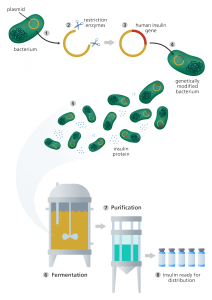
It has been more than 150 years since Gregor Mendel’s pea plant experiment demonstrated that ‘invisible factors’ determine the traits of organisms. It is doubtful that it would cross his mind that one day people could directly make changes to the “invisible factors”, which we call genes today, and alter these traits. Since the first transgenic Escherichia coli obtained antibiotic resistance in 1970s, tools have been developed to modify the genes of microbes, cell lines, plants, animals, and even human patients.

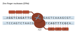
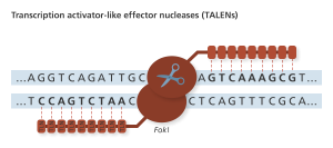
The principle of gene editing is based on the observation that cells can repair double strand breaks (DSB) through either the error prone pathway called non-homologous end joining (NHEJ) which sticks the two ends together or homologous recombination (HR) pathway that precisely repair the break using a template. Thus, the key to edit genes is to induce a site-specific DSB in the DNA sequence and allow the cell’s HR machinery to introduce the edit.

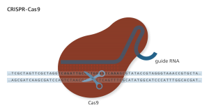
Standing out as a novel tool, the Clustered regularly interspaced short palindromic repeat (CRISPR)-CRISPR-associated protein 9 (Cas9) system was soon appreciated and widely used by biologists for gene editing since its discovery. Originally found in bacteria, CRISPR arrays contain short repeated DNA sequences separated by unique spacers acquired from phage DNA (protospacer) upon previous infection. When the same phage DNA is detected, the CRISPR array processes the corresponding CRISPR RNA (crRNA) and trans-activating CRISPR RNA (tracrRNA) that form a duplex to bring the nuclease Cas9 to the matching external protospacer DNA and destroy it. The phage DNA contains a 5’-NGG-3’ protospacer-adjacent motif (PAM) which is not present in the CRISPR array, so that the system can distinguish self-sequence and the phage DNA .

Given the ability to specifically cut the external phage DNA upon reinfection, the bacterial CRISPR/Cas9 system was soon adapted and developed as a tool for gene editing. CRISPR/Cas9 uses a single nuclease Cas9 and two RNA molecules to perform site-directed DNA editing. The crRNA identifies target DNA and the tracrRNA glues the crRNA to Cas9 to direct the protein to target DNA. Notably, a single guide RNA (gRNA) combines crRNA and tracrRNA into a single RNA molecule also has the full gene-editing activity in vivo and in vitro. Thus, a designed gRNA combines with Cas9 protein makes a versatile, cheap, easy to use, and efficient gene-editing tool.

Detailed structural biology studies has helped understand the complex mechanisms to this critical aspects of CRISPR/Cas9 action (reviewed by Jiang et. al. 2017). How does the Cas9/gRNA complex search the genome for the target? How is the nuclease activity of Cas9 only simulated at matching sequences? How prevalent are off-target effects, and how can the practitioner avoid these effects?


 How does the Cas9/gRNA complex search the genome for the target?

Cas9 is structurally organized into two distinct lobes, the target DNA recognition lobe (REC) that and the nuclease (NUC) lobe. The recognition lobe is made up of three alpha-helical domains, and the NUC lobe contains HNH-like and RuvC-like nuclease domains that create a break on each strand, as well as the C-terminal domain (CTD), which contains the PAM recognition site. The two lobes are connected by an arginine-rich linker (figure 1). Three major regulatory steps occur to achieve the efficient yet specific cut.

  • First, the gRNA binds  Cas9 which leads to a large  conformational change to activate Cas9 (Figure 1).
  • Second, the Cas9-gRNA complex binds to the target DNA PAM. PAM binding melts the adjacent DNA and plays an important role in crRNA/target-DNA strand hybrid formation (Figure 2).
  • Third, the perfect match of crRNA and the target strand induces another conformational change which activate the nuclease activity to make the double strand break  (Figure 3).

gRNA loading is the key for CRISPR to work. Without gRNA binding, the Cas9 protein is inactive and binds DNA weakly and nonspecifically. Structural studies also revealed  there is a large conformational change in Cas9 where Helix-III (Hel-III) in the REC domain moves towards the HNH nuclease domain upon guide RNA loading, illustrated in the Figure 1 cartoon.

Cas9/gRNA complex recognizes a 20-nucleotide (nt) complementary DNA sequence adjacent to the 5’-NGG-3’ PAM. With the conformational change, the 5’-NGG-3’ PAM recognition sites in the CTD domains are also repositioned to be able to form base-specific hydrogen bonds with the conserved GG (Figure 1). Moreover, the ribose-phosphate backbone of the guide RNA contacts with Cas9 and the 3’ 10-nt crRNA sequence (seed sequence) is prepositioned in a target binding favorable form. Now, the Cas9/gRNA complex is ready for target search.


Figure 1: Cas9 protein contains the nuclease (NUC) lobe and the recognition lobe (REC) that are connected by an  arginine-rich linker. Two arginines in the CTD (C-terminal domain)are exposed upon guide RNA(gRNA) loading, which will search for the target DNA PAM site and bind to it.  

Single molecule studies have revealed that Cas9/gRNA searches its target in a three-dimensional way rather than sliding on the DNA sequence. PAM probing is the key step to direct the Cas9 to potential targets. The base-specific hydrogen bonds between the GG and the R1333 and R1335 in the CTD domain are made in the major groove of DNA, which confers higher sequence specificity. A single mutation in the PAM site to 5’-NCG-3’ abolishes the double strand break created by CRISPR/Cas9. Interestingly, Cas9 with engineered (T1337R) PAM recognition site  engages a fourth guanine could recognize 5’-NGNG-3’ to introduce the DSB, further supports the importance of PAM recognition by CRISPR/Cas9.


How is the nuclease activity of Cas9 only simulated at matching sequences?

PAM-binding destabilizes the DNA duplex and triggers the Watson-Crick base pair between the crRNA and the target DNA (Figure 2). Next a series of three dynamic conformational changes occur to favor the DNA/crRNA hybrid formation, which is the rate-limiting step for Cas9/gRNA to make the double strand break revealed by kinetic studies.

  • First, the hydrogen-bond association of PAM with the CTD domain results in longer binding time between the gRNA and target DNA, which enables the following RNA/DNA hybrid formation.
  • Second, the phosphate in the target strand adjacent to the 5’ end of the PAM undergoes an unfavorable kink turn, which is stabilized by a phosphate lock loop (K1107-S1109) in Cas9. This kink contributes to both DNA double helix unwinding and the crRNA/DNA duplex formation.
  • Third, Cas9 CTD-domain makes Van der Waals interactions with the DNA phosphate backbone of the PAM-containing non-target strand. The nucleotide immediately upstream of PAM in the non-target strand stacks on the PAM duplex to stabilize it and the non-target strand kinks. The disordered non-target strand is then stabilized by the interaction between Cas9 and the -2 and -3 nucleotides. The kink in the non-target strand also helps to expose the two seed nucleotide strand to initiate RNA/DNA duplex formation. This is consistent with the observation that the mismatch of the two-crRNA nucleotide adjacent to PAM is intolerable while off-target DSB could occur in the DNA sequence homology to the seed region.


Figure 2: PAM-binding of Cas9/gRNA complex and initiation of crRNA/target DNA hybridization.

Upon PAM recognition and DNA/RNA duplex formation, each DNA strand is positioned to be cleaved by the nuclease domains HNH (target strand) and RuvC (non-target strand) 3nt upstream of the PAM. Off-target analysis suggested that there is far more Cas9/gRNA-DNA-binding events than the cleavage in the cell, which indicates that the PAM-binding alone is not enough to trigger the nuclease activity. In fact, 10-14nt complementary crRNA to the target strand is essential for the activation of the nuclease activity.

FRET experiments revealed that the distal complementary crRNA/target DNA duplex is a checkpoint which triggers a large conformational change to activate the HNH nuclease activity (Figure 3). The two hinge regions that link HNH and RuvC are important for RuvC nuclease activation to cut the non-target 3nt upstream of the NGG. Interestingly, the conformational change where HNH nuclease domain interacts with Helix-II (Hel-II) upon dsDNA binding might play important roles in locking the HNH domain in an active state. After the cleavage, the Cas9 protein remains to bind with PAM, and the in vivo mechanism to remove Cas9 to facilitate further DNA repair remains unclear.


Figure 3: Activation of Cas9 HNH and Ruv-C nuclease activity to create the double strand break


How can the practitioner increase efficiency decrease off-target effects?

With the structural knowledge of how  CRISPR/Cas9 produces a location-specific DSB, people can control the process by protein engineering for better gene-editing or even broader applications. For example, point mutation on the nuclease domain HNH (H840A) and RuvC (D10A) converts the enzyme into nickase, which only creates a single strand break on non-target strand or the target stand, respectively. The advantage with a DNA nickase is DNA nicks tend to induce HR DNA repair pathway over the error prone NHEJ (Maizels and Davis, 2018). Interestingly, the cleavage efficiency of the paired engineered Cas9 (D10A) nickases is more efficient or comparable to individual Cas9, suggesting engineering Cas9 for specific functions is possible without loss of efficiency (Gopalappa et al., 2018). A recent study showed that fusion of Rad51 (a DNA repair protein) or its variants with Cas9(D10A) nickase promotes HR DNA-repair at the DNA nicks and decreased off-target effects in some cell lines (Rees et. al. 2019). Fusion of Cas9 with a structurally unstable protein domain such as dihydrofolate reductase (DHFR) lead to the degradation of the protein under normal conditions. With trimethoprim (TMP) application, DHFR-Cas9 can be stabilized in a short period of time for gene editing, and the TMP can be removed to limit Cas9 activity to reduce off-target effects.


What else can we do with CRISPR/Cas9?

The double mutation of H840A and D10A results in a catalytic dead enzyme dCas9, which still retains the target DNA binding ability. dCas9 is widely used in different applications by fusion with various active domains. When fused with cytidine deaminase, Cas9 can become a base editor; when fused with transcriptional activators and repressors, Cas9 can be used to specifically regulate gene expression. Fusion of cytosine DNA methyltransferase with dCas9 can confer the protein epigenetic regulation ability in a sequence specific manner. Also, fusion of a fluorescent protein with dCas9 has been used for imaging genomic loci in live cells.

The last decade has witnessed the discovery and improvement of CRISPR/Cas9 tool kit  for both gene editing and other amazing applications. Yet, there still remains plenty of room for engineering more efficiency and precision to CRISPR/Cas9 function. It will be exciting to see how lessons and techniques learned in the CRISPR/Cas9 community lead to the next big thing in gene-editing. Let’s keep our eyes open to see what is going to come with the CRISPR/Cas9 legend!


References:

Jiang, Fuguo, and Jennifer A. Doudna. “CRISPR–Cas9 structures and mechanisms.” Annual review of biophysics 46 (2017): 505-529.

Gopalappa, Ramu, Bharathi Suresh, Suresh Ramakrishna, and Hyongbum Kim. “Paired D10A Cas9 nickases are sometimes more efficient than individual nucleases for gene disruption.” Nucleic acids research 46, no. 12 (2018): e71-e71.

Gong, Shanzhong, Helen Hong Yu, Kenneth A. Johnson, and David W. Taylor. “DNA unwinding is the primary determinant of CRISPR-Cas9 activity.” Cell reports 22, no. 2 (2018): 359-371.

Ribeiro, Lucas F., Liliane FC Ribeiro, Matheus Q. Barreto, and Richard J. Ward. “Protein engineering strategies to expand CRISPR-Cas9 applications.” International journal of genomics 2018 (2018).

Maizels, Nancy, and Luther Davis. “Initiation of homologous recombination at DNA nicks.” Nucleic acids research 46, no. 14 (2018): 6962-6973.

Rees, Holly A., Wei-Hsi Yeh, and David R. Liu. “Development of hRad51–Cas9 nickase fusions that mediate HDR without double-stranded breaks.” Nature communications 10, no. 1 (2019): 2212.

Graduate School Beyond the Lab

Author: Sharon Lee

Editor: Anna Feitzinger

How often have you looked through the window and thought to yourself, “Oh, it’s such a nice day outside. The sun is shining …” and immediately remember that you had signed up to use the microscope and will be inside a dark room for the rest of your afternoon?

Or wondered what else could you be doing during your graduate school years, other than meticulously pipet precise amounts of solutions and stare at computer screens to make sense of your fresh new data obtained using a protocol that took you half a year to optimize?

As graduate students, we are often so focused in completing our core classes, passing our qualifying exam, and gathering data for a publication that we forget all the other opportunities available to us in graduate school. We immerse ourselves into becoming the next Einstein that our days start and end at our lab doors.

Conducting rigorous research is definitely at the core of a successful graduate school experience. However, one might argue that gaining leadership and communication skills are just as important. So, whether you are simply looking for a reason to take a break and step outside or wanting something more to do beyond just your lab, check out some of the awesome graduate student groups on campus and consider volunteering for one of their fun events!

Young Scientist Program (YSP)

The Young Scientist Program is a collaborative effort between scientists and teachers to improve K-12 science education through interactive activities in the classrooms. Founded by BMCDB graduate student, Briana Rocha-Gregg back in 2014 and currently led by BMCDB graduate students Jennifer Baily and Abby Primack, YSP aims to empower all children to pursue higher education and careers in STEM fields. As graduate students, there are multiple ways to get involved in YSP from program management and event planning to designing fun instructional science worksheets for teachers to use in their classroom.

Jennifer and Aron Judd, a new YSP volunteer, joined the program as it reminded them about similar outreach work they did as undergraduates in their previous institutions.

 “Participating in YSP exposes you to the economic status of a lot [of] schools around Davis and puts you on the forefront of inspiring children to pursue science.” – Aron Judd Mendiola, current YSP volunteer and 1st year BMCDB graduate student.

For George Bell, a veteran YSP member since 2015, it was the students’ enthusiasm for playing with microscopes and extracting strawberry DNA that he found infectious and a worthy goal to pursue.

If you are passionate about giving back to the community and serving those who may not have access to science education, YSP can provide you with the opportunity to make a difference! Check out their website and look out for upcoming volunteering events.

STEM for Girls

STEM for Girls is an annual one-day outreach event hosted by the UC Davis Women’s Resource and Research Center in collaboration with Associate Professor Dr. Tina Jeoh. Organized by a committee of graduate students from various graduate groups, STEM for Girls invites over 50 middle school girls from the Woodland and Sacramento areas to UC Davis for a full day of interactive immersion into STEM. The goals of the program is to build the confidence of these young children, coming from mostly underrepresented and lower socioeconomic communities, in their abilities to participate in STEM and more importantly to introduce them to relatable and accessible STEM role models.

A Biomedical Engineering graduate student and a member in this year’s STEM for Girls planning committee, Alena Casella is excited to connect with the girls and be a role model for them. Although organized by graduate students, the STEM for Girls event is open to anyone at UC Davis who is interested in helping out as one of several team leaders or a volunteer. Undergraduate Charlyn Ritchie, who was a team leader last year, led a group of 10 girls to different workshops, lab tours and demonstrations through the day while answering any questions the girls had.

The 8th Annual STEM for Girls event will take place on Saturday, May 11th, 2019. Fill out an application form if you would like to volunteer by Friday, May 3rd, and share all things you love about science to a little one. If you missed this year’s deadline to volunteer, fret not and keep an eye out for the event when it comes back again next year!

Equity in Science, Technology, Engineering, Math and Entrepreneurship (ESTEME)

Formed by two graduate students, Nicole Nunez and Jeni Lee, originally as Women in Leadership back in 2013, ESTEME has come a long way since then. Today, the mission of this graduate student-run organization is to raise awareness and promote inclusion of all individuals interested in STEM and entrepreneurship. ESTEME is unique in its two branch systems where one branch focuses on outreach and the other in professional development. Current ESTEME members can take advantage of both and participate in either depending on their interests.

Under their Outreach branch head by Co-Vice Presidents, the organization provides graduate students with the opportunities to be involved in science communication and outreach at the K-8 level.

 “We both wanted to join a community that was actively engaged in improving diversity and addressing some of the inequities in STEM fields outside of the university setting.” – Alexus Roberts and Hannah Nelson, Co-Vice Presidents of ESTEME Outreach.

In the Professional Development branch, led by Vice President Linda Ma, ESTEME aims to bridge the gap for graduate students from underrepresented groups in the sciences to pursue STEM careers after their PhD.

Sign up to receive updates on when the next ESTEME outreach or professional development meeting will be taking place via esteme.ucd@gmail.com. Meanwhile, browse through their website to learn more.

Science Says

Science Says is a science communication group at UC Davis primarily made up of graduate students from various backgrounds and early career scientists. The overarching goal of the organization is to make science interesting, relevant, and accessible to everyone through communicating science in easy, comprehensible ways to the general public.

One of the ways the group achieves this goal is through their blog, Science REALLY says, which seeks to ensure that scientific data is accurately represented and not lost in translation when it reaches the broader audience through the media. Science Says also collaborates with other science communication groups such as CapSciComm from Sacramento and invites experts from the field of science communication to campus to train interested students.

Destiny Davis, current President of Science Says, found the organization to provide her an outlet for a different sort of creativity other than her research work, including a supportive community of scientists. Sydney Wyatt, Social Media Chair on the leadership team, joined the group out of her interest in written communication and to help the group’s mission to curate a well-informed scientifically literate public.

For more information about this dynamic group, visit their website and send them an email at ucdavisspcg@gmail.com if you are interested in becoming more involved.

BioScope

Stemmed from the idea and eagerness to share experiences of biomedical sciences graduate students, BioScope is a blog created by a group of BMCDB graduate students with the guidance of Professor Dr. Sean Burgess.

The online publication features written content covering topics ranging from science policy and ethical issues surrounding the use of scientific technologies to open discussions and tips for success in graduate school. Since its inception in 2018, BioScope has expanded to include students from other graduate programs. It also offers anyone in the UC Davis community with the opportunity to write special featured articles as invited writers without long-term commitments and involvement within the organization.  

Current Co-Editor in Chief and founding member, Keith Fraga describes BioScope as a collaborative opportunity for graduate students across the life sciences at UC Davis. BioScope is an up-and-coming graduate student group looking for new contributors to take on leadership roles and shape the organization’s future as it continues to grow.

 “Part of the challenge is balancing leadership roles with other graduate school responsibilities, but the outcome is always fulfilling.” – Anna Feitzinger, Co-Editor in Chief of BioScope.

Take a break and read one of the many articles published on the BioScope website. If you have an idea or a story in mind that you would like to share and write about, reach out to the group at UCDBioScope@gmail.com.


What can you do in an hour?

Author: Aiyana Emigh

Editor: Keith Fraga 


Earlier last month, the White House released the president’s FY2020 budget proposal. For those of us supported by non-defense federal funding sources, this proposal should worry you: the budget asks for a $54 billion (9%) drop in spending for R&D programs. A detailed analysis of the proposed R&D budget can be found here. However, there is hope.

This past week, I was selected by the UC Davis Government and Community Relations office to be one of two students sponsored by UC Davis to attend the annual AAAS CASE Workshop in Washington DC. This program included three days of workshops on science policy, advocacy and communication followed by a day of meetings with the offices of congressional members.


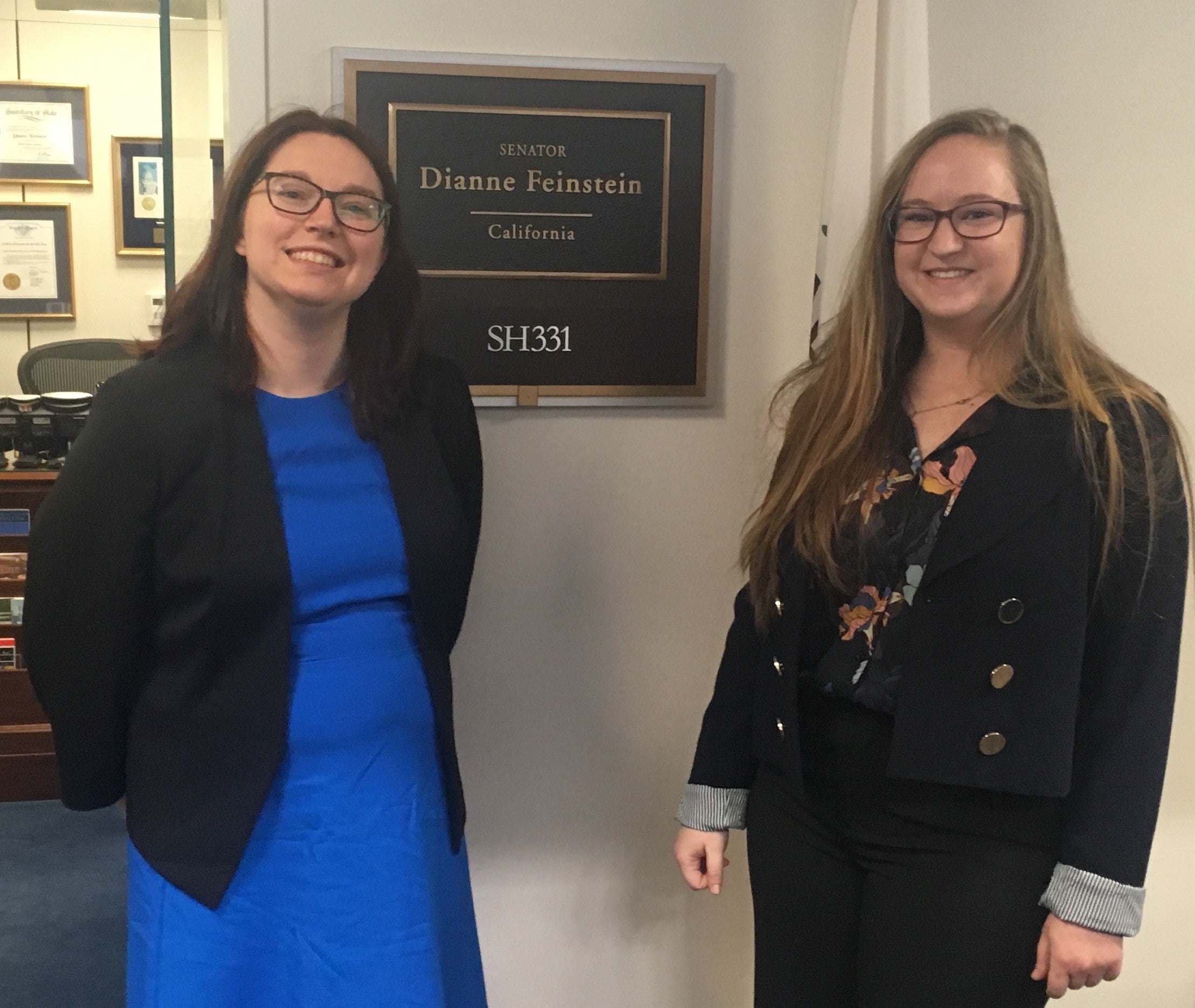
On our day of meetings, the California student delegation met with the offices of Senator Feinstein, Senator Harris, Speaker Pelosi, and House Minority Leader McCarthy. The UC Davis students additionally met with Representatives Garamendi, Bera, and Matsui who represent the greater Sacramento area. Every office expressed their fervent support for our research efforts.  While #MakingOurCASE for federal science funding, a legislative staffer in Pelosi’s office directly stated that the president’s budget was “not a starting point for negotiations.” There is strong bipartisan support for science funding.


However, this doesn’t mean we can sit back and relax. Although general support for science is strong, research on key politicized issues (such as climate change) is still controversial. And, with the Democrat majority in the House for the first time in 8 years, the funding of many important social programs are high priority and means a tighter budget. So the next question is: how can you get involved?


One of the most important workshop sessions I attended this week was led by Erin Heath, the Associate Director of Government Relations at AAAS. What I found significant during her talk was her recognition that graduate students are extremely busy–we don’t have a lot of time to spare and there is often an energetic barrier to trying unfamiliar things. She broke down her presentation into segments of what we could do in an hour, day, week, year or lifetime to participate in science advocacy efforts. In just one hour, you can:


  • Vote: This opportunity may only come up every once in a while, but it is one of the most important things you can do: help elect future leaders who are responsive to the needs of our community and will advocate on our behalf. It is easier than ever before to be an informed voter.


  • Learn: Do you not feel informed on a topic? Are you unsure who your representatives are? Do you want to know what is going on in the science policy world? Spend a free hour to research a topic, sign up for updates from science policy news sources, watch a webinar, discuss issues with people in your community, or check out resources available through your scientific society. The first barrier to action is lack of information.


  • Reach Out: This is the crucial time of year for science advocacy efforts. The Senate and House have just started holding appropriations hearings that will decide next year’s funding levels. Reaching out to your representatives and senators and telling them your story and why science funding is important to you and your district can be very powerful. This can be done with a quick phone call, email, or visit to their local offices. Alternatively (or additionally), you can meet with someone from our government relations office at UC Davis to share your story and/or talk to them about how to get involved.


The ability to make a difference is within reach. Stay informed, speak out, and take action.


Keep an eye out for more upcoming posts on the topics of science policy, advocacy, and communication and what you can do with more than one hour!


Don’t Fear the CRISPR

Author: Sydney Wyatt

Editor: Keith Fraga

Human genetic modification strikes fear into many scientists and non-scientists alike. The recent claim of human genetic editing experiments suggested that a researcher in China, Dr. He Jiankui, edited human embryos to be resistant to HIV, and that some embryos were successfully carried to term (1). While the twin girls, and a potential third baby on the way from another couple, seem to be healthy, this may not be the first time Chinese researchers have genetically modified humans (2).

An RNA guide targets the Cas9 nuclease through complementary binding. The variable guide length allows for relatively easy design with high specificity to the target site. Genome Research Limited

Since the advent of CRISPR*, a specific genetic editing tool derived from a native bacteria defense system, genetic modification is the new hot ticket for research and media coverage (3). The technology is so accessible, citizen-scientists popularly known as biohackers have attempted to modify themselves with DIY CRISPR kits (4). While self-experimentation is discouraged, it begs the question: should scientists genetically modify a human using CRISPR?

Process of using recombinant DNA to engineer E. coli to produce human insulin for mass production. Genome Research Limited

Genetic editing is not a new technology. Recombinant DNA** has existed since the 1970s and is used extensively in research and biotechnology companies to create genetically modified organisms (5). For example, Genentech and Eli Lilly & Co used recombinant DNA to genetically modify bacteria in order to mass produce human insulin used to manage diabetes (approved by the FDA in 1982). Genetically modifying bacteria dramatically increased yield and purity of insulin over animal-sourced insulin, improving the access to this vital therapeutic for insulin patients.

At the time of its discovery, there was concern within the science community as well as within the general public over gene manipulation in humans using recombinant DNA. In February 1975, biologists, lawyers, and journalists gathered for the second Asilomar Conference on Recombinant DNA to draft regulations on experiments using recombinant DNA technology (6). Just prior to the conference, a moratorium on research projects using recombinant DNA had been voluntarily put in place and universally observed — a remarkable example of scientists’ ability to self-regulate (7). The recommendations that emerged from the conference addressed “how the scientific work could be undertaken with minimal risks to workers in laboratories, to the public at large, and to the animal and plant species sharing our ecosystems.”

Ultimately, the safety precautions were laid out in guidelines issued by the NIH in July 1976 but never became law despite legislators’ suggestions. Researchers instead continued self-regulation with the added guidance of the NIH Recombinant DNA Advisory Committee. It seemed like excessive red tape to deal with in order to perform any experiments using this technology, but the regulations were necessary to ensure public trust in scientific endeavors (7). In the decades since, these regulations have been refined to address new concerns in the field while maintaining forward momentum.

The far-reaching effects of the 1975 Asilomar Conference remains unmatched today. The new tech on the block is CRISPR, which is a far more nuanced and powerful genetic editing tool than recombinant DNA.

CRISPR/Cas9, commonly referred to as CRISPR in the media, is an engineered nuclease. And it’s not the first. Meganucleases (c. 1980s), zinc finger nucleases (ZFN; c. 1990s), and transcription activator-like effector nucleases (TALEN; c. 2010) make up the larger family of engineered nucleases. ZFNs, TALENs, and CRISPR (c. 2013) have been used successfully for genetic editing (8).

Zinc fingers (ZF) recognize codons, or 3-base codes in the DNA, to guide the FokI nuclease to the cut site. Genome Research Limited

ZFNs were the first truly programmable genetic editing tool and was applied in humans to disrupt the CCR5 co-receptor that HIV uses to enter cells. Preclinical trials using ex vivo somatic cell gene editing successfully demonstrated that using ZFN gene therapy for HIV treatment was feasible and safe and thus merited clinical trials with the intent to take this treatment to market (9). Phase I and Phase II clinical trials are being conducted in the United States regarding the treatment of HIV patients; clinical trials are also underway for several other diseases (10). Importantly, these trials edit only somatic cells of the patient, not the germline cells (eggs or sperm), so the changes made will not be inherited. I spoke with gene therapy researcher Dr. David Segal of University of California, Davis, about these clinical trials.

“The things that go into clinical trial, by government regulation, have to pass certain thresholds for safety and efficacy…We strive to say anything that goes into a person is as safe as we know it to be. So everyone should know [that] about clinical trials. They go to great lengths to demonstrate safety above all else – even before efficacy – when you go into clinical trial.” – Dr. David Segal

TLN sites recognize single bases, allowing for more freedom in choosing targets for editing with the FokI nuclease. Genome Research Limited

TALENs were next on the scene, but were quickly outshined by CRISPR. Thus, human genetic editing using TALENs has not made it to clinical trials in the United States.

This brings us full circle to CRISPR’d humans. It is interesting that He Jiankui, the man behind the CRISPR babies, also chose to target CCR5. Since the original MIT Technology Review article was published, a January 21 report from Xinhua, China’s state media agency, has confirmed the birth of genetically edited twin girls and the pregnancy of another couple, suggesting a third genetically edited could be born (11).

Was this ethical? According to the report, no. Segal elaborated on the ethical measures we have regarding genetic editing in humans and that He bypassed these ethical measures for his germline editing experiments:

“[W]hen science starts to push up against ethical boundaries, we have institutions in place to try to respect, society’s concerns about the research that are being done…it’s absolutely essential that all the work go through this committee…anything you do that involves people, even if it’s a psychological questionnaire that at the worst case could cost someone some mental harm, all the way up to working with human embryos…has to be approved by an institutional review board. And again, they want to maintain transparency…They are the oversight at this institution. They have different people on the committee. There’s kinds of protections that they look for. All the people doing the research need to have training in ethics of human research that involves the unethical behaviors that had been done in the past and how we need to avoid that. If we don’t train the investigators, if we don’t go through the IRB, if we don’t follow this procedures, these institutions that have been set up to maintain transparency and trust and the scientific endeavor in the eyes of society, the investigator could lose his job. The NIH can stop funding the entire university. I mean, there are big consequences [in] trying to circumvent these structures that have been put in place to maintain society’s confidence in the scientific ventures. And I would say that was the most egregious thing that scientist in China did.”

He Jiankui blatantly broke these agreements, resulting in the ethical uproar over his experiments. Segal also discussed the long-term consequences of germline gene editing and gene therapy in our interview, which can be read in full here.

Interestingly, a group of scientists, including a few of the original discoverers of CRISPR and recombinant DNA, issued a statement published in Science in 2015 requesting a moratorium on human genetic modification (12). Echos of the recommendations from the Asilomar Conference are present in the recommendations laid out in the statement. According to Segal, some of these recommendations have come to pass – CRISPR research transparency is highly encouraged through the establishment of forums like the International Summit on Human Genome Editing where He unveiled his work.

The recommendations: 1) Discourage, even in countries that might be permissive, any research aimed at heritable genetic modification; 2) Establish forums for information and education on the risks and rewards of using CRISPR to treat or cure human disease, and the accompanying ethical, social, and legal concerns; 3) Support research transparency to help determine whether or which clinical applications are permissible; 4) Gather experts in genetics, law, and bioethics as well as members of the scientific community and public at large to consider the issues at hand.

A publication from 2017 discussed similar concerns about whether we are prepared for CRISPR clinical trials (13). The publication mentions a Phase I clinical trial was already in progress in China with the intention of treating stage IV metastatic non-small cell lung cancer, but that a similar trial was still prospective in the United States. The authors offered an extensive and critical review of the preclinical data that was presented to the NIH Recombinant DNA Advisory Committee (sound familiar? It’s the same committee that was established around the time of the Asilomar Conference).

In short, they determined we are not ready. This conclusion was informed by an existing framework for assessing the jump from preclinical to clinical trials (14-17).

Yet on February 1, NPR published an article on human genetic editing that is happening in the United States (18). Dieter Egli of Columbia University claims that he is conducting his experiments genuinely for research, in contrast to He’s supposed goal of genetically protecting the babies from HIV. Currently, Egli is focused on correcting one of the underlying genetic defects that result in inherited blindness and only allows the modified embryos to develop for one day, though he hopes to allow further development if these initial experiments are successful.

Federal funds are banned from being used for this kind of research – germline editing – in the United States, but there is no such stipulation on private funding (i.e. self-funded, as implied in the case of He’s work). If you recall, DIY CRISPR kits are readily available online, so if one has the means, then there is nothing preventing DIY designer babies beyond one’s ethics. There is still considerable controversy: should the moratorium be instilled? Should it be applied to basic research, such as Egli’s project? Can adequate regulations be established to prevent the need for a moratorium?

According to Segal, “we’re in the very early days of trying to use this as a therapy. Most of the work doesn’t involve any humans, but some things are progressing to a point where it can be used in humans, and in clinical trial.”

Only time will tell. In the meantime, don’t fear the CRISPR. It won’t be coming to a human near you anytime soon.

Thank you Dr. Segal for taking the time to provide his expert opinion on this topic.


*CRISPR: a genetic engineering tool using a short, repetitive DNA sequence and associated editing protein Cas9 to specifically edit target DNA sequence.

**Recombinant DNA: DNA made by artificially combining DNA fragments from different organisms.


For more history on genetics, check out The Gene by Siddhartha Mukherjee.

Bibliography:


  1. Regalado, A. (2018, November 26). EXCLUSIVE: Chinese scientists are creating CRISPR babies.
  2. Foley, K. E. (2018, January 26). Chinese scientists used Crispr gene editing on 86 human patients.
  3. Doudna, J. (2015, January). Genome Engineering with CRISPR-Cas9: Birth of a Breakthrough Technology.
  4. Lee, S. M. (2019, January 17). This Biohacker Is Trying To Edit His Own DNA And Wants You To Join Him.
  5. Herbert W. Boyer and Stanley N. Cohen. (2017, December 11).
  6. Berg, P., Baltimore, D., Brenner, S., Roblin, R. O., III, & Singer, M. F. (1975). Summary Statement of the Asilomar Conference on Recombinant DNA Molecules. PNAS, 72(6), 1981-1984. doi:10.1073/pnas.72.6.1981
  7. The Paul Berg Papers: Recombinant DNA Technologies and Researchers’ Responsibilities, 1973-1980. (n.d.).
  8. Chandrasegaran, S., & Carroll, D. (2016). Origins of Programmable Nucleases for Genome Engineering. Journal of Molecular Biology, 428(5), 963-989. doi:10.1016/j.jmb.2015.10.014
  9. DiGiusto, D. L., Cannon, P. M., Holmes, M. C., Li, L., Rao, A., Wang, J., . . . Zaia, J. A. (2016). Preclinical development and qualification of ZFN-mediated CCR5 disruption in human hematopoietic stem/progenitor cells. Molecular Therapy — Methods & Clinical Development, 3. doi:10.1038/mtm.2016.67
  10. Search of: Zfn | United States – List Results. (n.d.).
  11. Cross, R. (2019, January 21). Rogue CRISPR scientist will be punished. C&EN97(3), 1-56.
  12. David, B., Berg, P., Botchan, M., Carroll, D., Charo, R. A., Church, G., . . . Yamamoto, K. R. (2015). A prudent path forward for genomic engineering and germline gene modification. Science,348(6230), 36-38. doi:10.1126/science.aab1028
  13. Baylis, F., & McLeod, M. (2017). First-in-human Phase 1 CRISPR Gene Editing Cancer Trials: Are We Ready? Current Gene Therapy, 17, 309-319. doi:10.2174/1566523217666171121165935
  14. Kimmelman J. (2009). Gene transfer and the ethics of first-in-human research: lost in translation. Cambridge University Press.
  15. Henderson V.C., Kimmelman J., Fergusson D., Grimshaw J.M., & Hackam D.G. (2013). Threats to validity in the design and conduct of preclinical efficacy studies: A systematic review of guidelines for in vivo animal experiments. PLoS Med, 10(7):e1001489.
  16. Kimmelman J., & Henderson V. (2015). Assessing risk/benefit for trials using preclinical evidence: a proposal. J. Med. Ethics, 42(1), 50.
  17. Kimmelman J., & London A.J. (2011). Predicting harms and benefits in translational trials: ethics, evidence, and uncertainty. PLoS Med, 8(3):e1001010.
  18. Stein, R. (2019, February 01). New U.S. Experiments Aim To Create Gene-Edited Human Embryos. Retrieved from https://www.npr.org/sections/health-shots/2019/02/01/689623550/new-u-s-experiments-aim-to-create-gene-edited-human-embryos
fruit flies on marula fruit

Fruit Flies’ All Time Favorite Fruit

Author: Anna Feitzinger

Editor: Sharon Lee

Undeniably, you have seen a flurry of little fruit flies whizzing around left out bananas or oranges on a kitchen table. This little pest has been co-living, or commensal, with humans for thousands of years. But almost all of Drosophila melanogaster’s closest cousins, with the exception of Drosophila simulans, don’t enjoy human company (1)  – so how did this specific species become so chummy with man? Researchers from Lund University have published their discoveries (2) of previously unknown aspects of the natural ecology of one of the longest standing model organisms, which may explain how D. melanogaster became a human commensal and spread across the world.

Like humans, D. melanogaster has its origins in Africa. D. melanogaster migrated out of Africa and began to colonize Europe and Asia an estimated 10,000 years ago (3) . Given the commensal nature of D. melanogaster, it is no surprise that it has become one of the most widely used model organisms. Despite it’s annoyance around ripened fruit, the chromosomal theory of inheritance, genetic control of early development, and fundamental principles in neurobiology, population and evolutionary genetics all have a basis in D. melanogaster research. Given the century-long use of D. melanogaster in the laboratory, it is surprising that few fundamental aspects of the natural ecology of D. melanogaster is known. Interactions of organisms with their environment drive adaptation, shaping their evolution and inevitably  the present day biology that we study. It has been said that D. melanogaster lives where it eats, and for the first time we now know the likely ancestral host of D. melanogaster: the marula fruit.

Suzan Mansourian and colleagues set out to Matopos National Park in Zimbabwe to determine the host fruit of wild South African populations of D. melanogaster. The extremely abundant marula fruit has a similar pH to an orange, a highly fermentable pulp and contains terpenes and esters which are known olfactory cues for D. melanogaster. These characteristics, paired with the fact that domesticated D. melanogaster’s favorite breeding substrate is the orange, made the marula fruit the perfect candidate. Indeed, wild D. melanogaster were found in fly traps containing marula placed on the forest floor.

To test the hypothesis that flies from native habitats prefer marula to other fruit, the researchers placed paired traps containing either marula or orange under fruiting marula trees. D. melanogaster showed a strong preference for the marula fruit, although their sister species D. simulans, also native to the region, did not. Traps containing marula placed in locations with no fruiting marula trees, but other fruiting trees, caught none or very few D. melanogaster. These experiments have garnered D. melanogaster a new title: seasonal specialists!

How do domesticated flies react to marula fruit? Canton-S strain flies, originally collected sometime before 1916 in Canton, Ohio, prefer marula when given the choice between orange and marula. This conserved preference is impressive given that marula is not found outside sub-saharan Africa. To determine what specific chemical mediates this preference, the researchers tested major chemical components in a two-choice assay and found a high preference of esters responsible. Furthermore, using functional imaging of transgenic flies, the primary marula ester ethyl isovalerate was shown to activate Or22a-expressing olfactory sensory neurons (ab3a/b). In contrast, odor from oranges triggered weak to no activity. Silencing of the Or22a pathway using RNAi reduced the ability of flies to localize to marula compared to controls, suggesting that this pathway is responsible for detection and localization to marula.

If the Or22a circuit is linked to the specific chemistry of host fruit, Mansourian et al reasoned that local adaptation of the Or22a receptor would be found in fly populations from environments which contain different hosts. To investigate this, olfactory receptors sequences from 10 different African genomes and 1 European genome were analyzed. They found that Or22a and its adjacent paralog Or22b indeed showed genetic differentiation, a sign of local adaptation, between populations in contrast to other olfactory receptors. Next, they wanted to know if the sequence differences in Or22a and Or22b between populations confer functional changes. Measurements from ab3a neurons in a strain carrying a prevalent African variant of the receptor, Or22a/b (a fused gene as a result of a deletion), was found to be even more sensitive to the marula ester than non-African flies, confirming that the genetic differences are functional.

The now-vanished San tribes, known for their elaborate cave paintings, inhabited the Matopos during the Late Pleistocene to Early Holocene periods. Like D. melanogaster, the San appear to also have been seasonal specialists on marula – in one cave alone at least 24 million marula pits were recovered. This link may explain how D. melanogaster became a human commensal – wandering into San inhabited caves to feast on marula. Interestingly, traps placed inside the caves caught a number of D. melanogaster, but not D. simulans. The estimated date of the within-Africa expansion of D. melanogaster corresponds roughly with the date that marula harvesting ceased. D. melanogaster may have become dependent on marula harvesting by the San and when this ceased left the region with the San as a human commensal. Thus, the marula fruit may have been the glue that brought humans and one our most beloved model organisms into cohabitation, sparking their migration across the world and eventually into our present day laboratories.

Descendants of the ancient San have long been an interest to researchers. A 2010 paper in Nature showed that there is more genetic diversity between two San genomes than between an Asian and European genome (4). More recently, the San people became the first African group to draft a code of ethics for researchers who use their sequencing data (5). Although the use of the marula fruit by the ancient San ceased about ~10,000 years ago, it is still used commercially today both in beverages and cosmetics. Perhaps enjoying amarula, a cream liqueur made from marula, will help give a taste of what brought man and D. melanogaster together.

Author: Anna Feitzinger 


  1. Keller A (2007) Drosophila melanogaster’s history as a human commensal. Curr Biol 17(3):77–81.
  2. Mansourian S, et al. (2018) Wild African Drosophila melanogaster Are Seasonal Specialists on Marula Fruit. Curr Biol 28(24):3960–3968.e3.
  3.  Pool JE, et al. (2012) Population Genomics of Sub-Saharan Drosophila melanogaster: African Diversity and Non-African Admixture. PLoS Genet 8(12). doi:10.1371/journal.pgen.1003080.
  4. NordlingMar L (2017) San people of Africa draft code of ethics for researchers. Science | AAAS. Available at: http://www.sciencemag.org/news/2017/03/san-people-africa-draft-code-ethics-researchers [Accessed February 6, 2019].
  5. Schuster SC, et al. (2010) Complete Khoisan and Bantu genomes from southern Africa. Nature 463(7283):943–947.

San Code of Ethics available to read here: http://www.globalcodeofconduct.org/affiliated-codes/

I Work For You

Author: Keith Fraga

Editor: Sydney Wyatt


Current political turmoil in the US and abroad fosters uncertainty in the support for science. However, despite political and social divisions, science funding has experienced surprising support. Where does this support come from and where can scientists have an optimistic perspective of the future?

I argue the source of optimism for scientists is the long-standing positive relationship between science and the public. Broadly speaking, the relationship between science and the public consists of two different problems evaluated at two resolutions. On one end, the relationship is driven by passing policies and laws that advance scientists’ ability to do science. At this resolution, scientists can positively interact with citizens to influence public opinion and contribute to an informed public.

At the policy level, optimism comes from the historical and present bipartisan support for science funding. The support for science is one of the best examples of cooperation between Democratic and Republican lawmakers. As seen in Figure 1, government funding for science has exponentially increased since the1950s. This trend continues in 2018 where the federal science research budget increase was the largest in over 10 years.

As graduate students and scientists, we have a high demand for competitive science research funding from the federal government. In uncertain political times, we need to rely on the traditions of bipartisanship to continue the historical support for science.


[This image is directly from a Congressional Research Report written by John F Sargent Jr.]

Yet, there still remains significant political uncertainty in the future support for science. For instance, President Trump was reluctant to sign the 2018 spending bill that brought the substantial increase to US research funding, threatening the success of future deals. Future of science funding worldwide is uncertain due to rising populist governments, such as in Brazil’s President Jair Bolsonaro.

This is where scientists could focus on public opinion. Public opinion has a remarkable effect on government policy, proving the importance of maintaining our relationship with citizens. Fundamentally, bipartisan legislation advancing US research funding was undergirded by widespread public support for science.

The National Science Foundation sponsors surveys measuring public sentiment on science. They recently compiled a report demonstrating that public support for science continues to be high. Over 75% of Americans strongly approve of government supporting science research. Additionally, Americans maintain high confidence in the science community second only to the military.  

Public confidence in science is not without limits. On specific scientific problems, the public is skeptical of supporting issues like climate change, vaccination, and genetically modified food.   

Therefore, there still remains room for improvement in building more trust in scientific information. Furthermore, curating positive support from the public requires maintenance through effective communication.

This past September, Dr. Mary Woolley, the CEO of science policy interest group Research!America, discussed two strategies on how scientists can personally engage with citizens.

First, scientists understand that support for science pushes society into the future and can use a “Then-Now-Imagine” argument structure to describe the importance of support to non-experts.

For example, when curating support for vaccine development:  “Remember back then, polio was a major world-wide disease? Now, we don’t have to worry about polio because we developed a vaccine. Imagine if we can do that with cancer or HIV. That is what science can do.” This “Then-Now-Imagine” strategy can help citizens see the real return on investment in science.  

The second skill Dr. Woolley presented was the “I work for you” approach. Since the government has a major share of US science funding, scientists salaries come in whole or in part come from taxes. Scientists in many respects truly work for the citizens due to this fact.

So when a non-scientist asks you, “Hey, what do you do for a living?”, we can directly say, “Well, I actually work for you.” Dr. Woolley during her seminar shared two powerful anecdotes where individual scientists used the “I work for you” approach.

In one story, a scientist was at a coffee shop working, and out of curiosity from the interesting graphs on the scientist’s laptop, another customer asked what they do for a living. The scientist took the “I work for you” approach, and proceeded to share exactly how they worked for this curious citizen. The citizen was so impressed that they ended up endowing the scientist’s Department Chair position.

In a different setting, a scientist was on a plane, and struck up a conversation with the passenger next to them. Again, this scientist used the “I work for you” strategy, and the fellow passenger was inspired to organize a petition to support science funding.

These anecdotes don’t directly demonstrate the broad effectiveness of the “I work for you” strategy. But imagine, what if someone said that to you? How would you feel? Would you be curious? Would you want to learn more? It’s not everyday you randomly learn you have employees in this world. The key, ultimately, is we all can take initiative in positively impacting people we meet by explaining the productive investment they make in supporting science.  

Like many fellow graduate students, I struggle seeing the divisive ideas in American politics regarding science policy. At times, I focus too much on the political drama and feel pessimistic about the future. But in writing this article, speaking to peers, attending science advocacy seminars, I see an optimistic path to a fact-based future. I am optimistic, knowing that small interactions with fellow citizens could have surprising impacts. Alone, my impact may be small, but together we can have a powerful effect.


Further Reading

There is a wide, and complex literature on the effects of public opinion on government policy. I found the article Burstein P. (2010) Public Opinion, Public Policy, and Democracy. In: Leicht K.T., Jenkins J.C. (eds) Handbook of Politics. Handbooks of Sociology and Social Research. Springer, New York, NY to demonstrate utility of public opinion on formation of policy and specific issues with correctly evaluating public opinion’s impact. For the purposes of this article, public opinion impact on science funding is well documented.

BaMBA 2018 poster

A Day of Biology and Mathematics in the Bay Area

Saturday, November 3rd marked the 12th Annual Biology and Mathematics in the Bay Area Conference (BaMBA). BaMBA has developed into a particularly unique conference where you can expect high-level research and opportunities for interdisciplinary collaboration. BaMBA 2018 was held at the incredible Clark Center at Stanford University.

This year’s BaMBA meeting featured five fantastic experts presenting topics ranging from statistical methods in machine learning to cell biology of the immune system: Dr. Lacramioara Bintu (Stanford University)  Dr. Sean Collins (UC Davis), Dr. Bin Yu (UC Berkeley), Dr. Dexter Hadley (UC San Francisco), and Dr. Andrew Fire (Stanford University).


I had a chance to speak with Dr. Massa Shoura of Stanford University, an organizer for BaMBA 2018, about what makes BaMBA unique. “What I like about it (sic) brings people together that usually don’t go to the same conferences.” Interdisciplinary conferences like BaMBA are important opportunities for attendees to gain new perspectives on their research.


“As we grow older scientifically we end up specializing and then we end up going to the same conferences and talking to the same people because we are trying to solve something very intricate, but it is nice every now and then for us to get out of our comfort zone and talk to people who we usually dont talk and we find out this person can look at the same problem from a different angle and we end up having diversity in the way we solve the problem.” – Dr. Shoura


Dr. Shoura also pointed out that, because BaMBA is funded from several sources such as UC Davis College of Biological Sciences, the registration fee is waived for all attendees, allowing a broad audience of students, professors, and professionals from across the Bay Area to attend.


Diana Sernas, a 4th year undergraduate student at UCSC (‘19), also thought the accessibility of BaMBA was an important element to the conference’s uniqueness and success. According to her, “[a]ny opportunity for undergraduates to present their work is just another [opportunity for] professional development.” She also presented a research poster about her work on analyzing the 3D architecture of the genome with Dr. Javier Arsuaga.


“Presenting a poster at a conference is a great way for undergraduates to meet other people passionate about science and to see where they might fit in. “We are having an opportunity to look like a scientist, to be a scientist, to present ourselves as scientists.” – Diana Sernas (‘19)


I was especially excited for Dr. Sean Collins’ presentation from the UC Davis Department of Molecular & Cellular Biology. Dr. Collins’ research focuses on understanding the complex regulation of neutrophil chemotaxis vital in the immune response. With the use of live-cell reporters, the Collins lab is able to quantitatively interrogate how neutrophils locate pathogens in complex, noisy environments.


Dr. Collins also pointed out the significant impact BaMBA’s accessibility and well-organized schedule has had on its success: “I think it’s a really great meeting. It’s a nice scale [and] easy to interact with people [even] with a full day…  I have also been enjoying breadth of the meeting with really different talks and posters. It’s very nice.”


In addition to the main speakers, sessions/panels, and poster sessions, Q&A sessions offered time to discuss the material and, in some cases, resulted in initiating collaborative projects. Dr. Bin Yu of UC Berkeley “really enjoyed [BaMBA]” and was especially happy “to talk to students and [make] professional connections with other speakers.”


I highly recommend attending BaMBA in the future! It is an unique, accessible conference and an incredible opportunity to meet other passionate scientists. Keep an eye out for information about future BaMBA conferences at the the website: bambameeting.org. You can expect to see me there at BaMBA 13!


Author: Keith Fraga

Editor: Sydney Wyatt

Do Regulations on Genomic Data Inform or Mislead the Public?

Author: Emily Cartwright

Editor: Hongyan Hao


As sequencing costs drop and more companies develop genomic testing for everything from predicting hair color to the risk of disease susceptibility, the public has access to a wealth of personalized health information. At the same time, companies are faced with the decision of how much information to release to the public, only some of which is regulated. The Food and Drug Administration (FDA) has policies on what kinds of information that companies such as 23andMe and Ancestry.com can release to the public, but there are many enterprises that are not clinically certified and utilize genomic data to conduct health studies (1). How to regulate and disseminate findings from these companies, especially smaller scale studies that may not be clinically certified, is an unresolved issue.

Many of the current FDA regulations for companies like 23andMe are disease specific. Regulations released in 2017 allowed 23andMe to report on variants associated with 10 diseases or conditions (2) and in 2018, allowed the release of information on three BRCA1/BRCA2 variants to consumers who submitted for genetic testing (3). These specific BRCA1/2 variants are known to be associated with an increased risk of developing cancer but there are over 1,000 identified BRCA mutations (3). While these regulations limit the amount of information that consumers are provided, they also circumvent the current issue of how to relay health information to patients in a truthful and informative way; where it is made clear that the results of genetic testing indicate the known risk of developing a certain condition.

It is crucial that companies emphasize that not having a genetic marker associated with a disease or condition does not mean that the individual will not develop the condition. Current regulations from the FDA seek to limit what information can be released to the public but do not address how large companies can best convey health information to consumers. There are also gaps in the ability of agencies like the FDA to regulate all health information gleaned from genomic data, as many small companies do not fall under their regulatory jurisdiction (1).

Recently, the National Academies of Sciences, Engineering, and Medicine (NASEM) released a report (4) encouraging researchers to relate findings from biological data analysis back to people whose samples were used in the studies. The report is intended to increase the flow of information from scientists to the public, but a central issue still involves the question of how to convey information in a way that is both meaningful and accurate, in terms of whether or not a finding has medical significance

Genetic variants may indicate risk of disease susceptibility, but this is not necessarily causal and relaying such information to the public can be tricky (1). Researchers are also faced with an increased cost of funding for these studies because determining how to relay information to the public and executing this can result in additional expense (1). The NASEM recommended that researchers should plan out what information they will release from their studies prior to starting them, which may also help the researchers to plan out the added cost of relaying this information (1,4).

The flow of information from researchers to consumers is important but the fact that many studies are carried out by third parties also lies at the forefront of this issue. Companies that do not do the initial genomic sequencing and that may not have the clinical or scientific background to make the diagnoses they relay to consumers may give misinformation (1). The New York Times reported on a case where a doctor had sent his genomic data to a third party, Promethease, and was told that he had a variant known to be associated with Lynch syndrome, a disease that predisposes individuals to cancer early in life (5). The variant was associated with the disease but was not known to cause it, and more importantly, after sending out for genetic testing at a medical diagnostic firm, the doctor received results confirming that he did not actually have the mutation (5). The discrepancy in results brings up the issue of who should be allowed to handle genomic data and how should companies that do not fall under the umbrella of FDA regulations be treated (1). Right now, there may not be a good way to handle third party testing centers that have not been clinically certified, but consumers should be aware of the potential misinformation in results from these companies.

While there are still grey areas concerning the regulation of findings from studies that utilize genomic information, there is an effort to move towards defining what should be released and how. As researchers move towards utilizing this type of data for health and genetic studies, organizations such as NASEM are becoming more critical to help define what and how this information is treated and disseminated. There are many third-party companies that will not fall under guidelines set out by the NASEM but there is still a need to regulate how and what these companies can do with genomic data as well as how their information is released to the public (1). This can come from both sides, in conveying more accurate and meaningful data to consumers and in being more direct about the difference between association and causality in the relationship of variants to disease or condition.


Works Cited:

  1. Couzin-Frankel, J. (2018). If you give your DNA and tissues to science, should you get a peek at what they might contain? Science (New York, N.Y.). http://doi.org/10.1126/science.aau7323
  2. Administration, U. F. A. D. (2017). FDA allows marketing of first direct-to-consumer tests that provide genetic risk information for certain conditions.
  3. Administration, U. F. A. D. (2017). FDA authorizes, with special controls, direct-to-consumer test that reports three mutations in the BRCA breast cancer genes.
  4. National Academies of Sciences, Engineering, and Medicine, Health and Medicine Division, Board on Health Sciences Policy, Committee on the Return of Individual-Specific Research Results Generated in Research Laboratories, Downey, A. S., Busta, E. R., et al. (2018). Returning Individual Research Results to Participants: Guidance for a New Research Paradigm. http://doi.org/10.17226/25094
  5. Kolata, G. (2018, July 2). The Online Gene Test Finds a Dangerous Mutation. It May Well Be Wrong. The New York Times. Retreived from https://www.nytimes.com/2018/07/02/health/gene-testing-disease-nyt.html


Additional Links:

Spit is a podcast put out by 23andMe and iHeartRadio that features celebrity dialogue, highlighting some of the social issues surrounding genomic data.

On privacy issues surrounding genomic data: https://www.theverge.com/2018/8/1/17638680/genetic-data-privacy-consumer-rights-guidelines-23andme-ancestry

And in the news: https://www.nytimes.com/2018/10/18/opinion/sunday/dna-elizabeth-warren.html

More on the role of NASEM in regulating the release of information from genomic studies: https://www.biospace.com/article/national-academies-report-provide-patients-with-clinical-study-results/

A Video is Worth 1,000 Pictures

Still of HIV virus from The HIV Life Cycle.

Dr. Janet Iwasa from University of Utah gave a stellar keynote address on animating biology for the annual Molecular & Cellular Biology Training Program retreat on Friday, October 19. The major challenge for science communicators is to explain complex abstract concepts in a non-specialist manner. Whether it’s explaining the origin of life or how HIV infects the body, animation will revolutionize science communication, according to Dr. Iwasa. This form of visualization makes material tangible to a non-specialist audience and truly captivates their attention. Many students at the retreat were inspired to start our animation careers immediately, including myself. However, Dr. Iwasa offered sage advice — “Build yourself a comfortable little niche…Think about what skills you have now, and how those could be applied to a new discipline or area of research to create a unique niche.” Though this curbed my short-lived dream of becoming a world-famous biology animator, her material is freely available to download.


Author – Sydney Wyatt

Editor – Keith Fraga

The Art in Science and Science in Art

Author: Anna Feitzinger

Editors: Hongyan Hao, Sharon Lee, Keith Fraga


At first glance, science and art may appear to be two areas of human activity that could not be more distantly related. I’d like to dismantle this idea of scientists and artists and explore the qualities shared by both. Neon color tube racks and primary-colored pipette tip boxes are some of the basics that are scattered about the biologist’s benchtop. Biologists often visualize the microscopic world using dyes or fluorescent molecules and lasers of various wavelengths of light. The result is an image (or video) of colorful molecules whose arrangement depends on the particular cellular landscape and whose interpretation is up to the scientist. In fact, the first image that surfaces when searching “immunostaining” resembles that of a Paul Jackson Pollock painting (Figure 1).

Figure 1.  Left: Immunostained section of brain tumor in Wikipedia, The Free Encyclopedia. Right: Number 1, 1949 by Jackson Pollock. 

The history of science is full of art. Before cameras, scientists had to draw out their discoveries as lens’ became better at high levels of magnification. Inspired by the drawings in Robert Hooke’s Micrographia, the tradesman Antony van Leeuwenhoek learned how to grind lenses so to take up the hobby of microscopy. He made the most powerful lenses of the time and consequently discovered bacteria, protists, sperm cells, blood cells, rotifers and more before his death in 1723. [1] Although he had no formal higher education, he is now considered the Father of Microbiology.

Another unlikely scientist, Santiago Ramón y Cajal, was a rebellious and anti-authoritarian youngster who was said to have been imprisoned at the age of eleven in 1863 for destruction using a homemade cannon [2]. An avid painter, Santiago’s illustrations of brain and neural anatomy have become iconic. Considered the Father of Modern Neuroscience he postulated the neuron theory; the law of the dynamic polarization of the neuron and discovered the axonal growth cone.

Figure 2: A depiction of neurons in the cerebellum by Santiago Ramón y Cajal

Visual artists confined to physical media must discover the nature of their medium. Different techniques require different media and skills. For instance, one must decide the dilution factor of oil paint to linseed oil or acrylic paint to water in order to create a preferred texture and consistency. Screen printers and photographers must learn the properties of the light sensitive emulsions and films with which they work. Musicians become familiar with the sound waves they produce and modulate. Much like scientists mixing reagents in the lab, artists have an array of media used for experimentation.

Scientists and artists both have a need to share their perceptions of the universe which drives dedication, patience and persistence. Both have visions which they spend years perfecting and require creativity for success. Both get feedback from their peers and share their work with the community. Scientists share their work at conferences and artists do so in galleries or on stage. Occasionally, these two worlds collide. Last year marks the twenty year anniversary of the “Worm Art Show” which was started in 1997 by Ahna Skop at the International C. elegans conference. Works presented at the very first show included the C. elegans genome sandblasted onto a piece of driftwood and a wooden construction of the C.elegans vulva [3]. The American Society of Microbiology now hosts an annual Agar Art competition. This years first place piece, “The battle of winter and spring” features a stunning portrait of two contrasting figures made using colored microbes resistant to different antibiotics on a plate of agar.

Artists and scientists share a common thread historically, and each rely on one another in the modern world. Some recent collaborations include the turning of data into sound, called sonification. Mark Ballorad of Pennsylvania State University in State College has received two $50,000 grants for a project aiding marine biologists in translating data from the deep ocean into sound. Being able to listen to large data sets allows for an entirely different perspective that may not be captured by more conventional visualization methods. A recent publication by a music professor and chemical biologist explore how turning protein sequence and structural information into melodies can facilitate analysis.

Here at UC Davis the Department of Design hosts the LASER Series: conversations in art and science. Each LASER event has four presentations from four different disciplines as unrelated as possible. These talks are meant to facilitate interdisciplinary conversations between artists, scientists and generalists. The seminar is part of an international speaker series, all which are documented and accessible online. Another Art/Science initiative on campus is the Art/Science Fusion program which aims to bring creative energies from the arts and sciences to foster innovation.


[1] Kruif PD (1926) Microbe Hunters. Transactions of the American Microscopical Society 45(3):259.

[2] McMenemey, W. H., M.D. (1952). Section of Pathology (Vol. 46). Proceedings of the Royal Society of Medicine.

For updated information on the LASER series at UCD: https://www.facebook.com/laser.ucd

Additional articles on this topic:

https://www.forbes.com/sites/quora/2016/03/16/why-art-and-science-are-more-closely-related-than-you-think/

https://www.ebsco.com/blog/article/the-steamy-relationship-between-art-and-science

https://cosmosmagazine.com/society/when-arts-and-science-collide


Page 3 of 4

Powered by WordPress & Theme by Anders Norén